Abstract
Enterocytozoon bieneusi is an opportunistic microsporidian parasite with zoonotic potential that causes gastrointestinal illness in humans and animals. This study aimed to investigate the presence and genetic diversity of E. bieneusi from cats in Korea and to assess the potential public health risks associated with zoonotic genotypes. Among the 137 feline fecal samples, 4 (2.9%) were PCR-positive for E. bieneusi. In addition, 2 E. bieneusi genotypes were identified: Type IV, a known zoonotic genotype belonging to Group 1, and KCAT1, a novel genotype with zoonotic potential belonging to Group 1. This study is the first to report on these genotypes from cats in Korea, most of which were companion cats visiting veterinary clinics. Despite the low detection rate, the presence of zoonotic genotypes in companion cats is a potential public health concern because of the close physical interaction between cats and their human caregivers. These findings indicate the importance of routine monitoring and the molecular characterization of E. bieneusi in companion animals to comprehensively understand their zoonotic transmission patterns and to guide future risk assessments and preventive strategies.
-
Key words: Enterocytozoon bieneusi, zoonosis, genotype, cat
The phylum Microsporidia includes obligate unicellular parasites that infect various vertebrate and invertebrate hosts, with
Enterocytozoon bieneusi being the most common species found in humans [
1]. Over 90% of human microsporidiosis cases are due to
E. bieneusi [
2], which causes opportunistic infections, especially in immunocompromised patients, such as those with acquired immune deficiency syndrome and cancer, as well as those who have undergone organ transplantation [
3,
4].
E. bieneusi cannot perform metabolic processes outside the host cell, and it can only survive externally in the spore stage. Upon ingestion by a suitable host, the spore invades the intestinal cells, causing infection. Mature spores are excreted through feces, and the life cycle of
E. bieneusi continues through contact with infected animals or humans or via consumption of contaminated water or food in the environment [
5].
Enterocytozoon bieneusi spores are oval and are among the smallest Microsporidia (0.70–0.98 μm × 1.08–1.64 μm) [
5], making microscopic diagnosis difficult and requiring expertise. Therefore, PCR-based genotyping, which mainly targets the ribosomal internal transcribed spacer (ITS) region, is widely used to identify genetic characteristics and diversity [
6]. To date, >685
E. bieneusi genotypes are widely distributed across more than 11 genotypic groups. Group 1 being the largest includes many genotypes found in humans and animals [
1,
5,
7], such as genotypes D and Type IV, which are identified in several countries, including Korea [
6].
In Korea,
E. bieneusi has been studied in industrial animals (cattle, pigs, and horses) and in wild animals (bats, raccoon dogs, deer, leopard cats, wild boars, and wild deer) [
8–
10]. The
E. bieneusi genotypes that are confirmed in Korea include CAF1, CEbD, D, H, hores1, KBAT3, Peru11, PigEBITS3 to PigEBITS5, and Type IV (group 1); BEB8, CEbA, CEbF, I, J, KBAT1, KBAT2, and KBAT4 (group 2); and horse2 (group 6) [
6,
9,
11]. However, only few studies have investigated the prevalence of
E. bieneusi in Korea, with only 1 study focusing on cats living in shelters on Jeju Island [
11]. Jeju Island is located approximately 80 km from the southern coast of the Korean Peninsula. Given its geographical isolation and volcanic origins, the region exhibits a climate and natural environment that are markedly distinct from those of the mainland. The presence of zoonotic genotypes in companion cats could pose a public health concern because of the close interaction between companion cats and their human owners. Moreover, shelter cats may come into contact with the shelter staff during their temporary stay and with new owners during adoption. Therefore, this study aimed to assess the prevalence of
E. bieneusi in cats, including companion and shelter cats, in Korea and to determine its public health implications.
From January 2017 to November 2022, veterinarians collected 137 feline fecal samples from animal clinics and shelters across Korea. Fresh fecal samples were placed in individual tubes, transported in ice-packed boxes at 4°C, and transported to the laboratory for DNA extraction. Sample information (including age, sex, season, region, the presence or absence of diarrhea, and animal source) was recorded, with unclear or questionable data labeled as “unknown.” Age was categorized into 5 groups: kittens (<6 months), juniors (<2 years), adults (<10 years), seniors (≥10 years), and unknown age. The sampling region was divided into northern (Gangwon and Gyeonggi), central (South Chungcheong, North Chungcheong, North Gyeongsang, and North Jeolla), and southern (South Gyeongsang, South Jeolla, and Jeju) regions in accordance with the administrative district boundaries.
Fecal samples from cats were collected in accordance with the project guidelines by veterinarians at local, government-run veterinary institutes during monitoring, surveillance, treatment, or regular medical checkups after the receipt of oral consent from the cat owners. Fecal collection did not cause any harm to the animals and did not require ethical approval.
DNA was extracted using a QIAamp Fast DNA Stool Mini Kit (Qiagen, Hilden, Germany) in accordance with the manufacturer’s instructions. DNA quantity and quality were evaluated using the Infinite 200 PRO NanoQuant plate reader (Tecan, Mannedorf, Switzerland). In addition, PCR amplification was performed using AccuPower HotStart PCR Premix (Bioneer, Daejeon, Korea), and nested PCR was performed to amplify the
E. bieneusi ITS region for detection [
12,
13]. For the first round of PCR, ITSF1 (5′-GGTCATAGGGATGAAGAG-3′) and ITSR1 (5′-TTCGAGTTCTTTCGCGCTC-3′) were used as the forward and reverse primers, respectively. For the second round of PCR, ITSF2 (5′-GCTCTGAATATCTATGGCT-3′) and ITSR2 (5′-ATCGCCGACGGATCCAAGTG-3′) were used as the forward and reverse primers, respectively [
14]. PCR amplification was performed using Mastercycler Pro (Eppendorf, Hamburg, Germany) under the following conditions: initial denaturation at 95°C for 5 min, followed by 35 cycles of denaturation at 95°C for 30 sec, annealing at 55°C for 30 sec, and extension at 72°C for 30 sec, with a final extension at 75°C for 5 min.
Among the 137 collected specimens, 4 were positive for
E. bieneusi (
Table 1), with a prevalence of 2.9%. This value was lower than that reported for other animals, such as cats in Korea (Jeju Island, 3.8%) [
11], wild animals (45.2%), calves (16.9%), cattle (14.9%), and pigs (14.2%) [
8], with only bats and wild deer showing lower prevalences of 2.6% and 6.8%, respectively [
8,
10]. Based on these studies, the prevalence rate of
E. bieneusi varies according to animal type, sample size, and geographical region. This variation was also observed in previous research on cats [
1,
11,
15].
In this study, the prevalence of E. bieneusi was analyzed according to region, season, sex, age, presence of diarrhea, and animal source. For statistical analysis, the chi-square test was performed using the IBM SPSS Statistics version 26 (IBM Corp., Armonk, NY, USA), and the results indicated no significant risks.
When comparing by region, the prevalence of
E. bieneusi was 3.1% and 5.6% in the northern and central regions, respectively. With regard to season, the prevalence of
E. bieneusi was 6.3% in summer, 3.6% in autumn, and 2.1% in winter. In addition, the prevalence of
E. bieneusi was highest in females (3.4%), followed by males (2.8%) and those of unknown sex (2.3%). With regard to age, the prevalence of
E. bieneusi was 4.5% in kittens aged 6 months, 4.2% in juniors aged <2 years old, and 2.3% in cats of unknown age. The
E. bieneusi ITS region was not detected in diarrheal stools, but it occurred with a prevalence of 3.4% in normal stools. Finally, the prevalence of
E. bieneusi was 3.1% in animal samples collected from clinics and 2.6% in samples obtained from shelters (
Table 1). Given the limited number of samples tested in each region and season, the association between the prevalence of
E. bieneusi and specific geographical regions were not confirmed. Although the results were not statistically significant in this study, a previous study on the prevalence of
E. bieneusi in Korean water deer showed higher positivity in the southern region (9/68, 13.2%) than in the central (3/89, 3.4%) and northern (0/24) regions [
10]. In this study, differences in the prevalence of
E. bieneusi by age were not statistically significant. Previous studies also did not find any significant difference according to age, although some have reported conflicting results [
16,
17]. Therefore, infection is more closely related to individual differences than age [
17]. Some of the specimens collected at clinics may have been taken from stray cats that were temporarily sheltered at the clinics or from stray cats that had been adopted by new owners and brought in for treatment. Therefore, further research on the source of infection is necessary.
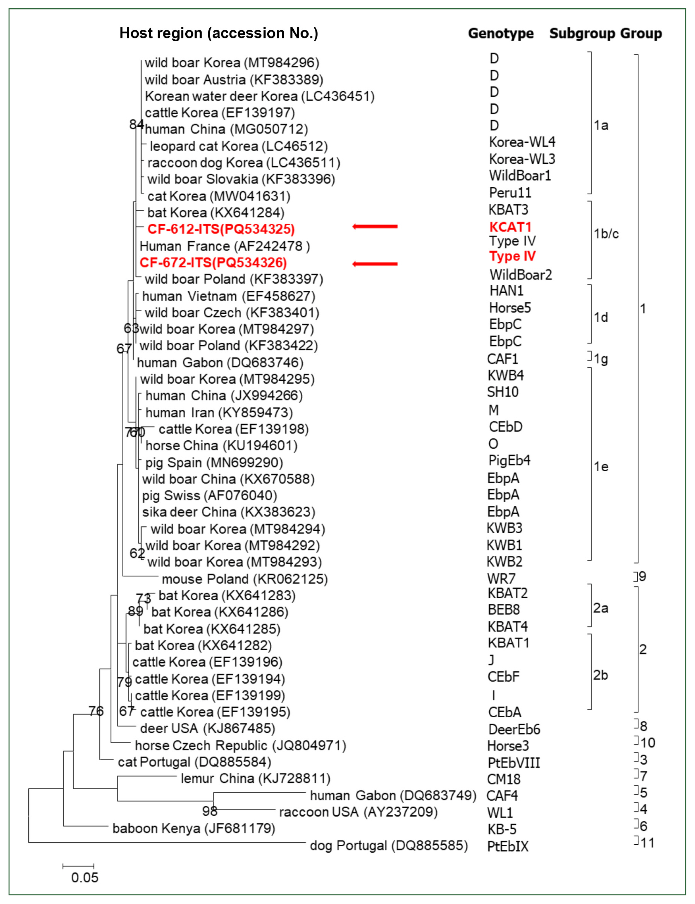
Four PCR-positive samples were sequenced at Macrogen (Daejeon, Korea), and 2 aligned sequences were obtained using BioEdit 7.2.5. Each sequence was submitted to GenBank (accession numbers PQ534325 and PQ534326). Phylogenetic analysis, including sequence comparisons via NCBI Web BLAST (
http://www.ncbi.nlm.nih.gov/blast), was also performed. The phylogenetic tree was constructed using MEGA7 [
18], and bootstrap analysis was performed using 1,000 replicates. All sequences belonged to group 1, a zoonotic group (
Fig. 1). Of the 4 sequences obtained, 3 were Type IV (PQ534326), and one was a new genotype, namely, KCAT1 (PQ534325). The new type, KCAT1, was isolated from a kitten that visited a clinic. The Type IV sequence obtained from humans (AF242478) was used as the reference sequence (243 bp) for genotypic comparison. The KCAT1 genotype exhibited 3 polymorphic sites at nucleotide positions 7 (T to G), 23 (C to T), and 64 (C to T). However, there were no mixed infections of
E. bieneusi genotypes were observed.
Group 1, the most diverse genotype, has several subgroups (1a–1i) and low host specificity. Along with Group 2, it is zoonoticand has been identified in various animals, including cats [
6]. More than 20
E. bieneusi genotypes, such as D, Type IV, Peru10, Peru11, and WL11, have been identified in cats and humans [
11]. In a previous study on
E. bieneusi from cats in Korea, only Peru11 (subgroup 1a) was identified [
11]. However, the genotypes identified in this study were type IV and KCAT1 genotypes, which are both classified as subgroup 1b/c of Group 1.
E. bieneusi genotype IV has been identified in humans and in a variety of animals, including cats, dogs, deer, and birds, indicating that this genotype is not restricted to a specific host and has a wide host range. On the contrary, the KCAT1 genotype differs from genotype IV by 3 nucleotides, indicating that it could be a potential transmission route for
E. bieneusi. However, further studies are necessary to investigate pathogenicity.
Type IV has been identified in cats in several countries, including Colombia, Germany, Portugal, Japan, China, and Türkiye [
6,
19]. A study in Australia identified a novel
E. bieneusi genotype in cats, namely, VIC_cat1 (MK696086), which differs from the zoonotic genotype IV by only 1 nucleotide [
17]. Type IV, which was previously unidentified in cats in Korea, has been confirmed in cow milk in Korea and is identical to that confirmed in humans (AF242478) [
20]. In addition, this Type IV genotype had a 100% sequence match with the human isolate (AF242478).
Dogs and cats are popular companion animals in Korea. They are commonly housed in homes or shelters, which increases their contact with people. In this study, the E. bieneusi sequences obtained from cats were 100% identical to those obtained from humans in other countries, indicating that cats represent a potential zoonotic reservoir.
This study is the first nationwide analysis of E. bieneusi from cats in Korea, including those from Jeju Island, covering shelter and companion cats visiting clinics. Genotype type IV, which has not been previously reported in cats in Korea, belongs to Group 1 and exhibits zoonotic potential, along with the new genotype KCAT1. Three of the 4 positive cases were identified in companion cats visiting veterinary clinics; thus, continuous monitoring and further research into the potential zoonotic transmission of E. bieneusi are warranted because of their close contact with humans. These findings underscore the importance of routine surveillance of E. bieneusi in companion animals. Preventive measures, including enhanced hygiene practices and regular fecal examinations targeting cat owners and veterinarians, are recommended. In addition, the molecular characterization of E. bieneusi is essential for a comprehensive understanding of its zoonotic potential, and it plays a critical role in future risk assessments and the development of effective prevention strategies.
Notes
-
Author contributions
Conceptualization: Kwak D
Data curation: Lee H, Kwak D
Formal analysis: Park HM
Investigation: Park HM, Lee H
Methodology: Park HM, Lee H, Sung SY
Project administration: Kwak D
Supervision: Kwak D
Validation: Nazim K, Jang BY, Sung KC, Lee SH, Seo MG, Rhee MH
Visualization: Park HM, Lee H, Nazim K
Writing- original draft: Park HM, Lee H, Sung SY
Writing- review and editing: Nazim K, Jang BY, Sung KC, Lee SH, Seo MG, Rhee MH, Kwak D
-
Conflict of interest
Dongmi Kwak serves as an editor of Parasites, Hosts and Diseases but had no involvement in the decision to publish this article. No other potential conflicts of interest relevant to this study were reported.
Fig. 1Analysis of Enterocytozoon bieneusi detected in cats in Korea based on the internal transcribed spacer region. The phylogenetic tree was constructed using the maximum likelihood method and bootstrap method with 1,000 replicates in MEGA7. The sequences identified in this study are indicated (arrows). Each sequence is described by host, country, and GenBank accession number. The genotypes (groups and subgroups) are also indicated on the right.

Table 1Prevalence of Enterocytozoon bieneusi in cats in Korea
Table 1
|
Variable |
|
No. of samples tested |
No. of samples positive for E. bieneusi (%) |
P-value |
|
Region |
Northern |
65 |
2 (3.1) |
0.626 |
|
Central |
36 |
2 (5.6) |
|
Southern |
31 |
0 |
|
Unknown |
5 |
0 |
|
|
Season |
Spring |
18 |
0 |
0.696 |
|
Summer |
16 |
1 (6.3) |
|
Autumn |
56 |
2 (3.6) |
|
Winter |
47 |
1 (2.1) |
|
|
Sex |
Female |
58 |
2 (3.4) |
1.000 |
|
Male |
36 |
1 (2.8) |
|
Unknown |
43 |
1 (2.3) |
|
|
Age |
Kitten (<6 months) |
44 |
2 (4.5) |
0.927 |
|
Junior (<2 years) |
24 |
1 (4.2) |
|
Adult (<10 years) |
22 |
0 |
|
Senior (>11 years) |
3 |
0 |
|
Unknown |
44 |
1 (2.3) |
|
|
Fecal type |
Diarrhea |
18 |
0 |
1.000 |
|
Normal |
119 |
4 (3.4) |
|
|
Animal source |
Clinic |
98 |
3 (3.1) |
1.000 |
|
Shelter |
39 |
1 (2.6) |
|
|
Total |
|
137 |
4 (2.9) |
|
References
- 1. Dashti A, Santín M, Cano L, de Lucio A, Bailo B, et al. Occurrence and genetic diversity of Enterocytozoon bieneusi (Microsporidia) in owned and sheltered dogs and cats in Northern Spain. Parasitol Res 2019;118(10):2979-2987. https://doi.org/10.1007/s00436-019-06428-1
- 2. Chen M, Wang H, Li X, Guo Y, Lu Y, et al. Molecular epidemiology of Enterocytozoon bieneusi from foxes and raccoon dogs in the Henan and Hebei provinces in China. BMC Vet Res 2024;20(1):53. https://doi.org/10.1186/s12917-024-03883-6
- 3. Liu X, Wu Y, Yang F, Gong B, Jiang Y, et al. Multilocus sequence typing of Enterocytozoon bieneusi isolates from various mammal and bird species and assessment of population structure and substructure. Front Microbiol 2020;11:1406. https://doi.org/10.3389/fmicb.2020.01406
- 4. Han B, Pan G, Weiss LM. Microsporidiosis in humans. Clin Microbiol Rev 2021;34(4):e0001020. https://doi.org/10.1128/CMR.00010-20
- 5. Nourrisson C, Lavergne RA, Moniot M, Morio F, Poirier P. Enterocytozoon bieneusi, a human pathogen. Emerg Microbes Infect 2024;13(1):2406276. https://doi.org/10.1080/22221751.2024.2406276
- 6. Li W, Feng Y, Santin M. Host secificity of Enterocytozoon bieneusi and public health implications. Trends Parasitol 2019;35(6):436-451. https://doi.org/10.1016/j.pt.2019.04.004
- 7. Ruan Y, Xu X, He Q, Li L, Guo J, et al. The largest meta-analysis on the global prevalence of microsporidia in mammals, avian and water provides insights into the epidemic features of these ubiquitous pathogens. Parasit Vectors 2021;14(1):186. https://doi.org/10.1186/s13071-021-04700-x
- 8. Lee H, Seo MG, Lee SH, Oem JK, Kim SH, et al. Distribution and genotypic analysis of Enterocytozoon bieneusi from wild boars in Korea. Med Mycol 2021;59(9):934-938. https://doi.org/10.1093/mmy/myab030
- 9. Lee H, Lee SH, Lee YR, Kim HY, Moon BY, et al. Enterocytozoon bieneusi genotypes and infections in the horses in Korea. Korean J Parasitol 2021;59(6):639-643. https://doi.org/10.3347/kjp.2021.59.6.639
- 10. Noh G, Lee H, Lee SH, Seo MG, Kim KT, et al. Genotypic analysis of zoonotic Enterocytozoon bieneusi in wild deer in Korea. Parasites Hosts Dis 2024;62(4):484-489. https://doi.org/10.3347/PHD.24072
- 11. Kwak D, Seo MG. Genetic analysis of zoonotic gastrointestinal protozoa and microsporidia in shelter cats in South Korea. Pathogens 2020;9(11):894. https://doi.org/10.3390/pathogens9110894
- 12. Feng S, Jia T, Huang J, Fan Y, Chang H, et al. Identification of Enterocytozoon bieneusi and Cryptosporidium spp. in farmed wild boars (Sus scrofa) in Beijing, China. Infect Genet Evol 2020;80:104231. https://doi.org/10.1016/j.meegid.2020.104231
- 13. Zhang XX, Cong W, Lou ZL, Ma JG, Zheng WB, et al. Prevalence, risk factors and multilocus genotyping of Enterocytozoon bieneusi in farmed foxes (Vulpes lagopus), Northern China. Parasit Vectors 2016;9:72. https://doi.org/10.1186/s13071-016-1356-1
- 14. Karim MR, Dong H, Li T, Yu F, Li D, et al. Predomination and new genotypes of Enterocytozoon bieneusi in captive nonhuman primates in zoos in China: high genetic diversity and zoonotic significance. PLoS One 2015;10(2):e0117991. https://doi.org/10.1371/journal.pone.0117991
- 15. Erkunt Alak S, Can H, Değirmenci Döşkaya A, Sürgeç E, Güvendi M, et al. Molecular prevalence of Enterocytozoon bieneusi in stray cats of Izmir, Turkiye. Comp Immunol Microbiol Infect Dis 2023;100:102037. https://doi.org/10.1016/j.cimid.2023.102037
- 16. Wang H, Lin X, Sun Y, Qi N, Lv M, et al. Occurrence, risk factors and genotypes of Enterocytozoon bieneusi in dogs and cats in Guangzhou, southern China: high genotype diversity and zoonotic concern. BMC Vet Res 2020;16(1):201. https://doi.org/10.1186/s12917-020-02421-4
- 17. Zhang Y, Koehler AV, Wang T, Cunliffe D, Gasser RB. Enterocytozoon bieneusi genotypes in cats and dogs in Victoria, Australia. BMC Microbiol 2019;19(1):183. https://doi.org/10.1186/s12866-019-1563-y
- 18. Kumar S, Stecher G, Tamura K. MEGA7: molecular evolutionary genetics analysis version 7.0 for bigger datasets. Mol Biol Evol 2016;33(7):1870-1874. https://doi.org/10.1093/molbev/msw054
- 19. Sürgeç E, Güvendi M, Karakavuk M, Erkunt Alak S, Değirmenci Döşkaya A, et al. Genotyping of Enterocytozoon bieneusi isolates detected in stray cats of Izmir, Turkiye. Parasitol Res 2023;122(11):2729-2735. https://doi.org/10.1007/s00436-023-07974-5
- 20. Lee JH. Molecular detection of Enterocytozoon bieneusi and identification of a potentially human-pathogenic genotype in milk. Appl Environ Microbiol 2008;74(5):1664-1666. https://doi.org/10.1128/AEM.02110-07

 , Haeseung Lee2