Three Echinostome Species from Wild Birds in the Republic of Korea
Article information
Abstract
Three echinostome species, i.e., Patagifer bilobus, Petasiger neocomense, and Saakotrema metatestis, are newly recorded in the trematode fauna of the Republic of Korea. They were recovered from 3 species of migratory birds (Platalea minor, Podiceps cristatus, and Egretta garzetta), which were donated by the Wildlife Center of Chungbuk (WCC) and the Conservation Genome Resource Bank for Korean Wildlife (CGRB). Only 1 P. bilobus specimen was recovered from the intestine of a black-faced spoonbill (P. minor), and characterized by the bilobed head crown with a deep dorsal incision and 54 collar spines. Twenty P. neocomense were recovered from the intestine of a great crested grebe (P. cristatus), and they had a well-developed head crown with 19 spines and 2 testes obliquely located at the posterior middle of the body. Total 70 S. metatestis were collected from the bursa of Fabricius of 1 little egret (E. garzetta). It is characterized by stout tegumental spines covered in the entire leaf-shaped body, posterior extension of the uterus, presence of the uroproct and a well-developed head crown with 12 pairs of collar spines on each side. By the present study, these 3 echinostome species are newly added to the trematode fauna in Korea.
INTRODUCTION
Echinostomes (=Family Echinostomatidae) are flukes that can be easily distinguished from other flukes thanks to their typical head collars as well as the presence of collar spines around the anterior part [1]. This trematode group is composed of 355 species and 6 subspecies in 50 genera, and they are parasites of birds, mammals, and humans [2]. In Korea, total 14 echinostome species in 7 genera have been reported: Acanthoparyphium marilae Yamaguti, 1934; Acanthoparyphium tyosenense Yamaguti, 1939; Echinochasmus japonicus Tanabe, 1926; Echinochasmus perfoliatus (Ratz, 1908) Dietz, 1909; Echinoparyphium recurvatum (Von Linstow, 1873) Dietz, 1909; Echinostoma cinetorchis Ando and Ozaki, 1923; Echinostoma gotoi Ando and Ozaki, 1923; Echinostoma hortense Asada, 1926; Echinostoma miyagawai Ishii, 1932; Echinostoma revolutum (Froelich, 1802) Looss, 1899; Euparyphium murinum Tubangui, 1931; Himasthla alincia Dietz, 1909; Himasthla kusasigi Yamaguti, 1939, and Stephanoprora sp. [3,4,5,6,7,8,9,10,11,12,13].
Echinostomes of birds have been investigated only in a few avian species in Korea. Total 8 species, i.e., A. marilae, A. tyosenense, E. japonicus, E. recurvatum, E. gotoi, E. miyagawai, E. revolutum, and H. kusasigi, have been reported as the avian echinostomes. Their hosts were recorded as follows: A. marilae from the black scoter, A. tyosenense from the ruddy turnstone, great knot, black-tailed gull, white-winged scoter, and black scoter, E. japonicus from the domestic duck and great egret, E. recurvatum from the mallard, spot-billed duck, and great crested grebe, E. gotoi from the mallard and spot-billed duck, E. miyagawai from the mallard, domestic duck, and spot-billed duck, E. revolutum from the falcated teal, mallard, spot-billed duck, and great scaup and H. kusasigi from the dulin [3,5,7,9,14,15,16].
The present report provides a detailed description of the morphological features and measurements of 3 so far unrecorded echinostome species in Korea: Patagifer bilobus (Rudolphi, 1819) Dietz, 1909; Petasiger neocomense Fuhrmann, 1927, and Saakotrema metatestis (Saakova, 1952) Skrjabin & Baschkirova, along with a review of those that were previously reported.
MATERIALS AND METHODS
Parasite specimens were recovered from the visceral organs of their hosts. Bird carcasses were donated by the Wildlife Center of Chungbuk (WCC) and the Conservation Genome Resource Bank for Korean Wildlife (CGRB). A black-faced spoonbill (Platalea minor), coded as CGRB12252, was collected from Yeocha-ri, Hwado-myeon, Ganghwa-gun, Incheon Metropolitan City, Korea in May 2010. A great crested grebe (Podiceps cristatus) was collected from a factory district in Cheongju-si, Chungcheongbuk-do, Korea in February 2011. The host animal had a fractured clavicle and hypothermia. Veterinarians of the WCC tried to save the bird but it eventually died. A little egret (Egretta garzetta, WCC no. 20120224) with severe fractures was found in Yeongdong-gun, Chungcheongbuk-do, Korea, in September 2013 and was necropsied immediately after death. Carcasses of 2 additional birds were donated to our laboratory for parasitological examinations and stored at -20℃.
Visceral organs of the hosts were dissected and washed with tap water. Parasitic worms were recovered by filtration through sieves (mesh size: 250 µm to 1 mm). Collected worms were flattened with a coverslip pressure and fixed with 10% neutral buffered formalin. The specimens were stained with Semichon's acetocarmine and were dehydrated through a graded alcohol series. After clearing with xylene, morphological characteristics of each specimen were observed with a light microscope with a reticle.
Examined specimens were deposited at the National Institute of Biological Resources (NIBR), Incheon Metropolitan City, Korea and the Parasite Resource Bank (PRB), Cheongju City, Chungcheongbuk-do, Korea.
RESULTS
Family Echinostomatidae Looss, 1899
Genus Patagifer Dietz, 1909
Patagifer bilobus (Rudolphi, 1819) Dietz, 1909
One specimen was recovered from the intestine of a black-faced spoonbill (Platalea minor, CGRB12252). NIBR specimen number is KOSPIV0000193866.
Description (n=1): A ribbon-shaped body with blunt end. The longitudinal muscle was well developed. Tegumental spines were absent. The body length was 12.3 mm and the width was 1.6 mm at the level of the uterus and 1.3 mm at the ventral sucker. The ratio between the forebody and body length was 12.2%. The head collar was large and well developed, bi-lobed, fan shaped, and with a deep incision. The collar was 937 µm in length and 1,415 µm in width. A total of 54 collar spines were present. Lateral spines were similar in size to the angle spines. The oral sucker was small, measured 283×370 µm. The pharynx was small, oval in shaped and located immediately behind the oral sucker, measured 253×224 µm. The esophagus was 341 µm, with an intestinal bifurcation anterior to the cirrus sac. The cecum ran through the lateral side of the body to the posterior extremity. The cirrus sac was elongate-oval in shape, measured 468×263 µm and was located anteriodorsally to the ventral sucker. The ventral sucker was large, well developed, and measured 1,190×995 µm. The uterus was filled with numerous eggs, loops intercaecal, and located between the ventral sucker and ovary. Eggs in the uterine loops (n=23) measured 95-105 (96±18)×52.5-57.5 (54±1) µm. The ovary was oval in shape, displaced by the uterine loops, and measured 175×335 µm. Mehlis' gland was oval in shape, located behind the ovary, and measured 458×292 µm. The vitelline follicles were located laterally on the body, between the ventral sucker and the posterior extremity and overlapped the cecum. The excretory vesicle and the pore were obscured. There were 2 testes, tandem: the anterior testis measured 1,035×332 µm and the posterior testis measured 1,308×332 µm (Fig. 1A-C).
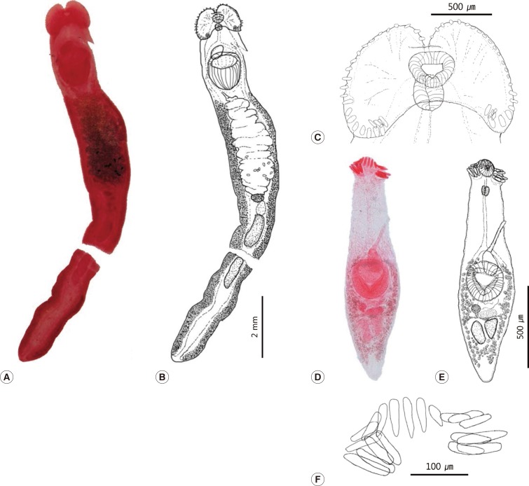
Light microscopic view of Patagifer bilobus (A-C) and Petasiger neocomense (D-F). (A) Photo of P. bilobus. (B) Drawing of P. bilobus. (C) Head part of the worm. (D) Photo of P. neocomense. (E) Drawing of P. neocomense. (F) Collar spines of the worm.
Genus Petasiger Dietz, 1909
Petasiger neocomense Fuhrmann, 1927
Total 20 specimens were recovered from the intestine of a great crested grebe (Podiceps cristatus). NIBR specimen number is KOSPIV0000192593.
Description (n=12): The body was fusiform, 1,191-1,470 (1,348±91) µm in length and 347-425 (395±26) µm in width at the level of the ventral sucker. It was covered with minute tegumental spines. The head collar was well developed, being 132-177 (145±14) µm in length, 213-272 (247±17) µm in width, and armed with 19 spines; 8 angle spines, 6 lateral spines and 5 dorsal spines. The angle spine was larger than the other spines, measuring 81-107 (93±7, n=37) µm long and 14-18 (15±1, n=37) µm wide, whereas the lateral spines were 53-61 (56±2, n=9) µm long and 8-13 (9±1, n=9) µm wide and the dorsal spines were 58-77 (67±5, n=16) µm long and 9-15 (12±1, n=16) µm wide. The oral sucker was subterminal, circular to oval in shape, 74-113 (88±12) µm long and 73-117 (88±13) µm wide. The prepharynx was 23-99 (55±20) µm long. The pharynx was oval, 65-79 (71±4) µm long and 49-61 (53±6) µm wide. The esophagus was 179-357 (262±55) µm in length. The intestinal bifurcation was anterior to the cirrus sac, which was muscular, located in the anteriodorsal region of the ventral sucker, and measured 120-181 (154±23) µm in length and 74-100 (84±8) µm in width. The cirrus, as observed in almost all specimens, was thin and long, measuring 17-30 (23±4) µm in width. The ventral sucker was large, located in the center of the body, measured 241-294 (274±18) µm long and 232-313 (281±26) µm wide. 2 testes were present, elongate-oval in shape, and positioned oblique or symmetrical. The anterior testis measured 107-181 (145±22) µm in length and 55-87 (71±10) µm in width, and the posterior testis measured 107-181 (145±22) µm in length and 49-87 (68±10) µm in width. The ovary was round, dextral, close to ventral sucker and measured 38-74 (54±10) µm in diameter. Mehlis' gland was elongate-oval, larger than ovary, 45-63 (57±6) µm long and 104-179 (133±22) µm wide. Vitelline follicles were only present in the hind body. The uterus was short and located behind the ventral sucker, and it contained only 1 or 2 eggs. The excretory vesicle was Y-shaped. Eggs were oval and measured 66-86 (73±7) µm long and 38-50 (43±4) µm wide (Fig. 1D-F).
Genus Saakotrema Skrjabin & Baschkirova, 1956
Saakotrema metatestis (Saakova, 1952) Skrjabin & Baschkirova, 1956
Total 70 specimens were collected from the bursa of Fabricius of 1 little egret (Egretta garzetta). Most of the worms recovered were damaged except for 3 intact specimens. NIBR specimen number is KOSPIV0000192596.
Description (n=3): A leaf-shaped body measuring 1.7-2.0 (1.86) mm long and 0.5-0.6 (0.55) mm wide, with the widest point at the level of the ventral sucker and narrowing at the posterior end. The entire body was covered with strong tegumental spines, 17 µm in length. The collar was well developed and measured 88-133 (115)×185-223 (203.3) µm. There were 24 collar spines in a single row. The oral sucker was well developed and measured 43-80 (66.7)×55-80 (68.3) µm. The pharynx was oval shaped, larger than oral sucker and measured 105-110 (108.3)×63-80 (70.0) µm. The intestinal bifurcation was located between the pharynx and the ventral sucker. Ventral sucker was large, well developed and measuring 278-300 (285.0)×260-303 (280.8) µm. The cirrus sac was obscure, oval shaped, and measured 135×75 µm (measurement taken from only 1 specimen). The ovary was oval in shape and measured 75-130 (108.3)×113-158 (132.5) µm. The testes were tandem, round to oval in shape, and were located immediately behind the ovary. The anterior testis measured 244-254 (240.6)×283-342 (306.0) µm and the posterior testis measured 322-332 (325.5)×234-322 (266.9) µm. The uterine coil was long and extended posteriorly through the ventral side of the worm. Eggs were oval and measured 70-83 (74.2±4.1)×43-48 (45.1±2.9) µm (Fig. 2A-D).

Light microscopic view of Saakotrema metatestis. (A) Photo of an adult worm. (B) Description of adult worm. (C, D) Collar spines of S. metatestis.
DISCUSSION
The present study revealed 3 unrecorded echinostome species in Korea. Since the first report of Echinostoma revolutum by Issiki [3], more than 14 echinostome species have been identified in Korea. Park [4] recovered Echinostoma hortense from rats, while Yamaguti [5] identified Acanthoparyphium marilae in Melanitta nigra, as well as A. tyosenense in M. nigra america and M. fusca stejnegeri as new species in Korea. E. cinetorchis and Euparyphium murinum were also recovered from wild rats by Seo et al. [6]. Chu et al. [7] reported the presence of E. revolutum, E. gotoi, E. miyagawai, and Echinoparyphium koizumi in wild birds. Echinochasmus perfoliatus was found from cats in Daegu-si [8]. Eom et al. [9] found E. miyagawai and E. japonicus in domestic ducks in Korea. However, E. miyagawai was considered as synonym of E. revolutum by Yamaguti [2,17] and Bykhovskaya-Pavlovskaya [18], but this species was re-validated upon closer examination of various characteristics, including the morphology and life cycle [19,20]. Lee et al. [11] found E. revolutum and Echinoparyphium recurvatum from house rats in Yangyang-gun, Gangwon-do and considered E. koizumi that reported by Chu et al. [7] as a synonym of E. recurvatum.
On the other hand, Himasthla kusasigi metacercariae were detected in the marine clam, Meretrix lusoria, and subsequently adults were recovered in the black-tailed gull, Larus crassirostris, experimentally infected in Korea [10]. This fluke was also recovered from the dunlin, Calidris alpina sakhalina, naturally infected in Ganghwa-gun, Gyeonggi-do [16]. Sohn and Chai [13] found E. revolutum, E. hortense, E. japonicus, and Stephanoprora in feral cats purchased in a market in Busan Metropolitan City. Metacercariae of Himasthla alincia were collected from an intermediate host, Mactra veneriformis, and a study was performed by infecting chicks [12]. However, the natural definitive hosts of this fluke have not been reported in Korea. Therefore, total 14 echinostome species were described in the Republic of Korea before the present study. Including the 3 new faunas in this study, a total of 17 species of 10 genera, i.e., Acanthoparyphium (2 species), Echinochasmus (2), Echinoparyphium (1), Echinostoma (5), Euparyphium (1), Himasthla (2), Petasiger (1), Patagifer (1), Saakotrema (1), and Stephanoprora (1), have been recorded to be distributed in Korea until now [6,7,8,9,10,11,12,13,14,15,16,21,22,23,24,25,26] (Table 1).

Definitive hosts of echinostomes other than humans in Korea
The worms recovered from Podiceps cristatus had 19 collar spines, including obvious longer angle spines, oblique to symmetrical testes, an elongate-oval cirrus sac and a tubular cirrus. These morphological characters were in line with those of P. neocomense, including size variation [27]. P. neocomense was first recovered from P. cristatus in Lake Neuchàtel by Furhmann [28]. In Korea, the host of P. neocomense is also P. cristatus. P. neocomense resembles P. nitidus morphologically but can be easily distinguished by the shape of the cirrus and the cirrus sac, as well as by geography (Eurasia vs North America) [27].
P. bilobus is the type-species of the genus Patagifer and was recovered from the Threskiornithidae birds in Europe, Asia, Africa, Australia, and America [29]. P. bilobus was the only known species found in spoonbills (genus Platalea) [29]. In this study, we recovered only 1 damaged specimen, which had similar characteristics to P. bilobus; a ribbon-like body, non-confluent vitelline fields, 4 pairs of angle spines on the ventral lappet and a deep dorsal incision on the head collar [29]. Our specimen was missing some of the tegumental spines and a section of the collar spines; however, we could confirm traces of these spines and identified the specimen as P. bilobus based on the morphology and host ranges. P. minor was added to the list as a new host of P. bilobus and was the first reported case of an echinostome fluke from the Threskiornithidae birds in Korea.
S. metatestis was first described by Saakova in 1952 and named Opisthotrema metatesti. However, in 1956, Skrjabin and Baschkirova [30] identified it as an echinostome species based on its echinostomid characteristics. They created a new genus, Saakotrema, for the fluke of Saakova and assigned it to the genus Saakotrema in the family Echinostomatidae [30]. The specimens collected in this study were identical morphologically (leaf-shaped body, posterior extension of the uterus, presence of the uroproct) and in size to S. metatestis (Table 2). S. metatestis has been recovered from piscivorous birds in Europe and Asia [31], including China [32]. This is, so far, the first report of S. metatestis in Far-East Asia.

Comparison of Saakotrema metatestis morphometrics with those of previous studies
Among echinostomes reported in Korea, 7 species (A. tyosenense, E. japonicus, E. perfoliatus, E. cinetorchis, E. hortense, E. revolutum, and E. recurvatum) were known as zoonotic parasites infecting humans [33]. There was no evidence that the 3 present species could cause human infections. However, in the case of A. tyosenense, it was primarily considered only as a parasite of birds [5], but 60 years later it was revealed to be a zoonotic parasite [34]. Thus, further monitoring is required in aspects of public health.
The Korean peninsula bridges China and Japan, so we can assume that helminth fauna overlaps the 2 countries. However, the investigation of parasitic fauna in Korea is not thorough enough. In China [32] and Japan, some faunistic revisions are available, but not in Korea. In this study, we found 3 unrecorded echinostome flukes from migratory birds in Korea and presented new area records for them. It is likely that many more unrecorded parasites are present in Korea, underlining the needs for additional studies.
ACKNOWLEDGMENTS
This work was supported by a grant from the National Institute of Biological Resources (NIBR), funded by the Ministry of Environment (MOE), the Republic of Korea (NIBR No. 2013-02-001). Parasite materials used in this study were provided by the Parasite Resource Bank of Korea, National Research Resource Center (2012-0000037), the Republic of Korea.
Notes
We have no conflict of interest related to this work.