Cross-reactivity between sera from dogs experimentally infected with Dirofilaria immitis and crude extract of Toxocara canis
Article information
Abstract
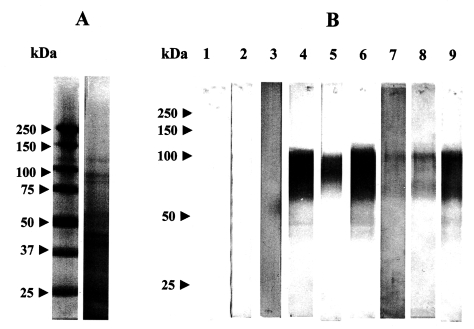
This study was performed to investigate whethere there is cross-reactivity between Dirofilaria immitis and three intestinal nematodes of dogs. In ELISA, D. immitis-infected dog sera obtained at the 4th molting stage (9-11 weeks) and microfilaremic stage (25-30 weeks) were shown to be highly reactive with crude extract of T. canis. In immunoblotting, some antigenic fractions, 44, 57, 88, 100 kDa of crude extract of T. canis, were found to be positive reaction with sera of dogs infected with D. immitis. However, little or no cross-reaction were observed between sera of D. immitis-infected dogs and crude extract antigen of T. vulpis or A. caninum. These result suggest that there are partial cross reaction between sera of D. immitis-infected dogs and the antigen of T. canis.
Employing the indirect hemagglutination assay (Hayasaki, 1981) and Dot-ELISA (Matsumura et al., 1984), the authors found no cross-reactivity was observed between Dirofilaria immitis and intestinal nematodes including T. canis. On the other hand, Ott et al. (1985) reported the presence of immunologically distinguishable species-specific non-cross reactive antigens and the presence of cross-reactive antigens in antigenic extracts obtained from adult D. immitis and T. canis. Additionally, Konno et al. (1995) reported that D. immitis possessed a partial antigenic cross reactivity to T. canis, revealed by using immunoblotting. The above consideration indicate that more strict analysis is required for the study of antigenic cross-reactivity between D. immitis and other parasites. This study was performed to examine on cross-reactivity between D. immitis and three intestinal nematodes of dogs.
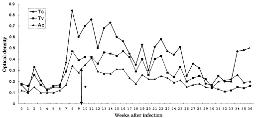
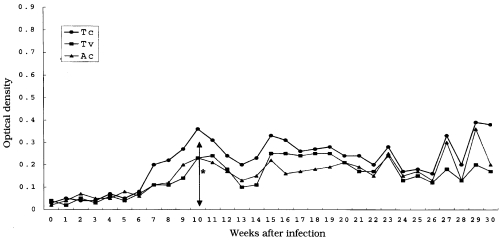
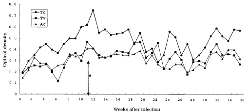
Five healthy mongrel juvenile dogs, approximately three months of age, were housed in mosquito-proof runs and treated with piperazine (100 mg/kg, PO) for intestinal parasites prior to experimentation. Three dogs were experimentally infected with D. immitis by the procedures described by Hayasaki (1982). Briefly, mosquitoes, Aedes togoi, collected during the larval stage from their natural spawning areas were reared under laboratory conditions and inoculated by feeding on a dog with about 200 circulating microfilarial (Mf) counts per 20 µl of blood. Two dogs were used as the negative control. Infective larvae (L3) of D. immitis were recovered from the proboscis of the infected mosquitoes 10 to 14 days after blood feeding and suspended in saline during microscopic observation. Three dogs were subcutaneously injected in the inguinal region with 228 (dog No. 1), 278 (dog No. 2) and 248 (dog No. 3) infective larvae (L3). The blood was collected from the cephalic vein once a week, and the sera were stored at -70℃ until analysis. The crude extract antigens of T. canis, T. vulpis and A. caninum were prepared as previously described (Hayasaki, 1982). Briefly, the crude antigens used in this study were extracted from adult worms of T. canis, A. caninum and T. vulpis by phosphate buffered saline (PBS, pH 7.2, 0.1 M). These worms were homogenized and sonicated by tissue homogenizer (15 min, 4℃) and ultrasonicator (50 watt, 15 min, 4℃), respectively, and then allowed to incubate overnight at 4℃. After centrifugation at 18,000 g, the supernatant as antigen was collected and kept at -70℃. The protein concentration of the antigens was determined using the methods of Lowry et al. (1951). ELISA was performed by method described by Grieve et al. (1981). SDS-PAGE and immunoblotting was performed as described by Gonno and Hayasaki (1995). In this study, by ELISA, D. immitis-infected sera obtained at the 4th molting stage (9-11 weeks after infection) and microfilaremic stage (25-30 weeks after infection) were strongly reacted with crude extract of T. canis. However, little or no cross-reactivities were found between D. immitis-infected sera and crude extracts of T. vulpis or A. caninum.
Immunoblotting analysis are more effective application of immunologic diagnosis (Kaneko et al., 1990; Hayasaki et al., 1994). In this study, 44, 57, 88 and 100 kDa, which were protein bands of crude extract of T. canis, were shown to be strongly reactive with D. immitis-infected sera. Based on these findings, we suggest that D. immitis share a common antigen with T. canis. This cross-reactivity may be dependent on many antigenic epitopes existing in somatic components of the parasites because previous report indicate that D. immitis consists of many protein components and its antigenicity is very complex, and also these two parasites may be originated from a same progenitor and still possess partially similar antigenicity. Therefore, certain interspecies antigenic epitopes may be isolated from one species which may be useful in eliciting a highly specific immune response in a host toward cross-reactive epitopes in different species (Hayasaki et al., 1994).
Further studies are needed to elucidate the immunological implications antigenic cross-reactivity between D. immitis and intestinal nematodes including T. canis.