Cho, Tak, Kweon, Kim, Choi, Kong, and Chung: A human case of Echinostoma hortense (Trematoda: Echinostomatidae) infection diagnosed by gastroduodenal endoscopy in Korea
Cited By
Citations to this article as recorded by
Evaluation of the role of rats as reservoir hosts for fishborne zoonotic trematodes in two endemic northern Vietnam fish farms
Nguyen Lan Anh Thi, Henry Madsen, Dao Thanh Ha, Eric Hoberg, Anders Dalsgaard, K. Darwin Murrell
Parasitology Research.2012; 111(3): 1045.     CrossRef
Biomphalaria straminea (Mollusca: Planorbidae) as an intermediate host of Drepanocephalus spp. (Trematoda: Echinostomatidae) in Brazil: a morphological and molecular study
Hudson A. Pinto, Matt J. Griffin, Sylvie M. Quiniou, Cynthia Ware, Alan L. Melo
Parasitology Research.2016; 115(1): 51.     CrossRef
Eosinophilia: Causes and pathobiology in persons with prior exposures in tropical areas with an emphasis on parasitic infections
Yae-Jean Kim, Thomas B. Nutman
Current Infectious Disease Reports.2006; 8(1): 43.     CrossRef
Epidemiological analysis of human fascioliasis in northeastern Punjab, Pakistan
Asma W. Qureshi, Akhtar Tanveer, Santiago Mas-Coma
Acta Tropica.2016; 156: 157.     CrossRef
Fasciola hepatica infection in children actively detected in a survey in rural areas of Mardan district, Khyber Pakhtunkhawa province, northern Pakistan
Asma W. Qureshi, Aurang Zeb, Abu Mansoor, Azam Hayat, Santiago Mas-Coma
Parasitology International.2019; 69: 39.     CrossRef
Neglected food-borne trematodiases: echinostomiasis and gastrodiscoidiasis
Rafael Toledo, María Álvarez-Izquierdo, J. Guillermo Esteban, Carla Muñoz-Antoli
Parasitology.2022; 149(10): 1319.     CrossRef
An update on human echinostomiasis
R. Toledo, J. G. Esteban
Transactions of The Royal Society of Tropical Medicine and Hygiene.2016; 110(1): 37.     CrossRef
ECHINOSTOMA HORTENSE ASADA INFECTION IN THE DUODENUM: INCIDENTAL FINDINGS DURING ROUTINE GASTROINTESTINAL ENDOSCOPY
Toshiaki Tanaka, Tomoari Kamada, Hideki Koga, Aki Tanaka, Noriaki Manabe, Jiro Hata, Hiroaki Kusunoki, Manabu Ishii, Motonori Sato, Kenichi Tarumi, Akiko Shiotani, Tetsuya Okino, Ken Haruma
Digestive Endoscopy.2008; 20(2): 87.     CrossRef
Human echinostomiasis: a case report
Ranjit Sah, Shusila Khadka, Rabin Hamal, Sagar Poudyal
BMC Research Notes.2018;[Epub]     CrossRef
Echinostoma hortense Infection with Enteritis Diagnosed by Upper Gastrointestinal Endoscopy in a Dog
Hiroki OKANISHI, Jun MATSUMOTO, Sadao NOGAMI, Yumiko KAGAWA, Toshihiro WATARI
Journal of Veterinary Medical Science.2013; 75(7): 991.     CrossRef
Rare Case of Echinostoma cinetorchis Infection, South Korea
Sooji Hong, Hyejoo Shin, Yoon-Hee Lee, Sung-Jong Hong, So-Ri Kim, Youn-Kyoung Kim, Young-Jin Son, Jeong-Gil Song, Jong-Yil Chai, Bong-Kwang Jung
Emerging Infectious Diseases.2024;[Epub]     CrossRef
A human infection of Echinostoma hortense in duodenal bulb diagnosed by endoscopy
Young-Doo Chang, Woon-Mok Sohn, Jae-Hwa Ryu, Shin-Yong Kang, Sung-Jong Hong
The Korean Journal of Parasitology.2005; 43(2): 57.     CrossRef
Echinostoma hortense and Heterophyid Metacercariae Encysted in Yellowfin Goby, Acanthogobius flavimanus, from Shinan-gun and Muan-gun (Jeollanam-do), Korea
Woon-Mok Sohn, Byoung-Kuk Na, Shin-Hyeong Cho
The Korean Journal of Parasitology.2009; 47(3): 307.     CrossRef
Foodborne Intestinal Flukes in Southeast Asia
Jong-Yil Chai, Eun-Hee Shin, Soon-Hyung Lee, Han-Jong Rim
The Korean Journal of Parasitology.2009; 47(Suppl): S69.     CrossRef
A Case of Echinostoma cinetorchis (Trematoda: Echinostomatidae) Infection Diagnosed by Colonoscopy
Woon Tae Jung, Kyeong Ju Lee, Hong Jun Kim, Tae Hyo Kim, Byoung-Kuk Na, Woon-Mok Sohn
The Korean Journal of Parasitology.2014; 52(3): 287.     CrossRef
Animal Models for Echinostoma malayanum Infection: Worm Recovery and Some Pathology
Jiraporn Songsri, Ratchadawan Aukkanimart, Thidarut Boonmars, Panaratana Ratanasuwan, Porntip Laummaunwai, Pranee Sriraj, Panupan Sripan
The Korean Journal of Parasitology.2016; 54(1): 47.     CrossRef
Complete Mitochondrial Genome of Echinostoma hortense (Digenea: Echinostomatidae)
Ze-Xuan Liu, Yan Zhang, Yu-Ting Liu, Qiao-Cheng Chang, Xin Su, Xue Fu, Dong-Mei Yue, Yuan Gao, Chun-Ren Wang
The Korean Journal of Parasitology.2016; 54(2): 173.     CrossRef
New Definitive Hosts and Differential Body Indices of Isthmiophora hortensis (Digenea: Echinostomatidae)
Woon-Mok Sohn, Byoung-Kuk Na, Sung-Shik Shin
The Korean Journal of Parasitology.2017; 55(3): 287.     CrossRef
Echinostoma macrorchis (Digenea: Echinostomatidae): Metacercariae in Cipangopaludina chinensis malleata Snails and Adults from Experimental Rats in Korea
Woon-Mok Sohn, Byoung-Kuk Na
The Korean Journal of Parasitology.2017; 55(5): 541.     CrossRef
Infection Status of Isthmiophora hortensis Metacercariae in Dark Sleepers, Odontobutis Species, from Some Water Systems of the Republic of Korea
Woon-Mok Sohn, Byoung-Kuk Na, Shin-Hyeong Cho, Jung-Won Ju
The Korean Journal of Parasitology.2018; 56(6): 633.     CrossRef
Morphology and Molecular Identification of Echinostoma revolutum and Echinostoma macrorchis in Freshwater Snails and Experimental Hamsters in Upper Northern Thailand
Preeyaporn Butboonchoo, Chalobol Wongsawad, Pheravut Wongsawad, Jong-Yil Chai
The Korean Journal of Parasitology.2020; 58(5): 499.     CrossRef
Clonorchis sinensis and Echinostoma hortense detected by endoscopy and molecular characterization: two case reports and update on diagnosis
Lijia Wen, Benhe Wang, Hui Zhang
Frontiers in Medicine.2025;[Epub]     CrossRef
Parasitic Diseases of Upper Gastrointestinal Tract
Heung Up Kim
The Korean Journal of Helicobacter and Upper Gastrointestinal Research.2024; 24(2): 127.     CrossRef

Abstract

A human Echinostoma hortense infection was diagnosed by gastroduodenoscopy. An 81-year-old Korean male, living in Yeongcheon-shi, Gyeongsangbuk-do and with epigastric discomfort of several days duration, was subjected to upper gastrointestinal endoscopy. He was in the habit of eating fresh water fish. Two live worms were found in the duodenal bulb area and were removed using an endoscopic forcep. Based on their morphological characteristics, the worms were identified as E. hortense. The patient was treated with praziquantel 10 mg/kg as a single dose. The source of the infection in this case remains unclear, but the fresh water fish consumed, including the loach, may have been the source. This is the second case of E. hortense infection diagnosed by endoscopy in Korea.

Members of the family Echinostomatidae are zoonotic flukes, which infect birds and mammals including humans. Human infections by the echinostomatid flukes, i.e. Echinostoma hortense, E. cinetorchis, Echinochasmus japonicus, and Acanthoparyphium tyosenense, have been reported in Korea (Seo et al., 1983; Ryang et al., 1986; Ahn and Ryang, 1986). Of the four species, E. hortense is the most frequently reported to be a causative fluke (Chai and Lee, 2002). Endemic foci of E. hortense have been previously reported in Umsong-gun, Chungchongbuk-do (We, 1987) and Chongsong-gun, Kyongsangbuk-do (Lee et al., 1988).
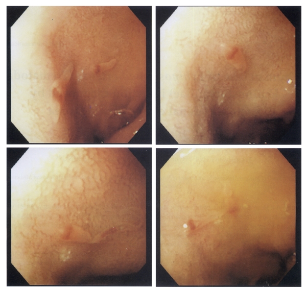
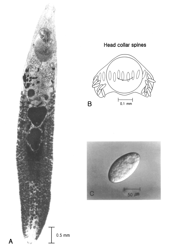
An 81-year-old Korean male, living in Yeongcheon-shi, Gyeongsangbuk-do, was admitted to the outpatient's department of Internal Medicine, Kyungpook National University Hospital. The patient complained of epigastric discomfort of several days duration. No significant abnormality was detected by laboratory findings. He was in the habit of eating raw fresh water fish. Gastroduodenal endoscopy revealed two live worms in the duodenal bulb area; the mucosa of which showed mild erosion (Fig. 1). The worms were picked out with an endoscopic biopsy forcep and transported to the Department of Parasitology, Kyungpook National University School of Medicine, where they were fixed in 70% ethanol and stained with Semichon's acetocarmine. They were very slender (7.92 × 1.33 mm), and had a characteristic head crown equipped with 27 collar spines around the subterminal oral sucker (Fig. 2A and B). The ventral sucker (0.68 × 0.77 mm) was positioned at the anterior 1/3 of the body and was much larger than the oral sucker (0.25 × 0.26 mm). The uterus was distended and contained thin-shelled elliptical eggs. A spontaneously discharged egg (124.3 × 68.6 µm) had an operculum at the narrower end and a well-defined germ ball (Fig. 2C). Two testes were distinct, slightly lobulated, and located in tandem at the equatorial portion. The worms were identified as E. hortense by morphological characteristics and measurements.
The symptoms of echinostomiasis vary from none to abdominal pain, although a case in Korea showed duodenal ulceration (Chai et al., 1994). Because of high incidence of stomach cancer and ulcer in Korea, many senior citizens with gastrointestinal symptoms are subjected to endoscopic examinations. Echinostoma hortense can be easily demonstrated by gastroduodenoscopy due to its habitat in the upper small intestine and its relatively large size. The present case is the second Korean case of E. hortense infection, which was diagnosed by gastroduodenoscopy (Chai et al., 1994). Four cases diagnosed by endoscopy have been reported in Japan (Hamamoto et al., 1997).
The source of the infection in this case is unclear. Many senior residents of inland areas of Gyeongsangbuk-do eat raw fresh water fish, including loaches, a known source of human infection. An epizootiological survey in the Yeongcheon area should be performed to determine the sources of human infection. The residence of this patient was Yeongcheon-shi neighboring Cheongsong-gun, which has been reported to be one of the endemic foci of E. hortense. Yeongcheon-shi and Cheongsong-gun are inlands of Gyeongsangbuk-do and contain upper reaches of the Nakdong-gang (river). The geographic characteristics of both localities coincide well with the indications of Chai and Lee (2002), namely, that E. hortense is distributed inland along the upper reaches of large rivers. Therefore, to confirm Yeongcheon-shi as an endemic area of E. hortense, an epidemiological survey of this fluke should be undertaken.

REFERENCES

1. Ahn YK, Ryang YS. Experimental and epidemiological studies on the life cycle of Echinostoma hortense Asada, 1926 (Trematoda: Echinostomatidae). Korean J Parasitol 1986;24:121-136.
crossref
2. Chai JY, Lee SH. Food-borne intestinal trematode infections in the Republic of Korea. Parasitol Int 2002;51:129-154. PMID: 12113752.
crossref
3. Chai JY, Hong ST, Lee SH, Lee GC, Min YI. A case of echinostomiasis with ulcerative lesions in the duodenum. Korean J Parasitol 1994;32:201-204. PMID: 7953246.
crossref
4. Hamamoto T, Kawasaki H, Maejima J, Hirai K. A case of Echinostoma hortense infection diagnosed by the upper gastrointestinal endoscopy. Nippon Shokakibyo Gakkai Zasshi 1997;94:487-491. PMID: 9277113.

5. Lee SK, Chung NS, Ko IH, et al. An epidemiological survey of Echinostoma hortense infectin in Chongsong-gun, Kyongbuk Province. Korean J Parasitol 1988;26:199-206.
crossref
6. Ryang YS, Ahn YK, Kim WT, Shin KC, Lee KW, Kim TS. Two cases of human infection by Echinostoma cinetorchis. Korean J Parasitol 1986;24:71-76.
crossref
7. Seo BS, Hong ST, Chai JY, Lee SH. Studies on intestinal trematodes in Korea VIII. A human case of Echinostoma hortense infection. Korean J Parasitol 1983;21:219-223.
crossref
8. We JS. Prevalence of intestinal helminthiasies in some residents at Umsong-gun, Chungchongbuk-do, and detection of metacercariae from fresh water fish. 1987, Seoul National University. Dissertation for Master of Science (MSc) in Medicine.

Fig. 1
Endoscopic images of two live parasitic worms in the patient's duodenal bulb.
kjp-41-117-g001
Fig. 2
Ventral view of an adult Echinostoma hortense stained with Semichon's acetocarmine (A), drawing of the head collar spines (B), and an egg (C).
kjp-41-117-g002
1