Abstract
Schistosomiasis remains a major neglected tropical disease, affecting approximately 600 million individuals worldwide and accounting for nearly 500,000 deaths annually. The principal causative species—Schistosoma haematobium, S. mansoni, and S. japonicum—drive significant morbidity through hepatomegaly, splenomegaly, and progressive hepatic fibrosis. Praziquantel (PZQ) remains the cornerstone of treatment; however, its limited efficacy against immature worms and eggs, combined with concerns over emerging drug resistance, underscores the urgent need for novel therapeutic alternatives. This study investigated the anti-schistosomal potential of green-synthesized zinc oxide (ZnO) nanoparticles (NPs) in a murine model of S. mansoni infection, benchmarked against PZQ. ZnO NPs were fabricated using ginger extract via an eco-friendly green synthesis approach. Fifty male BALB/c mice were randomly assigned to five groups (n=10): normal control, infected untreated control, infected treated with PZQ alone, infected treated with ZnO NPs alone, and infected treated with a PZQ–ZnO NP combination. Parasitological, histopathological, and fibrosis assessments were subsequently performed. All treatment groups demonstrated significant reductions in worm burden and tissue egg counts relative to infected untreated controls. Histopathological examination of untreated infected mice revealed extensive chronic granulomatous inflammation, concentric perioval fibrosis, fibroblast proliferation, hepatocellular necrosis, hydropic degeneration, marked collagen deposition, and portal-to-portal fibrous bridging. Treated groups, by contrast, exhibited marked hepatic improvement characterized by reduced granuloma size, diminished fibrosis, and decreased collagen deposition. Collectively, these findings indicate that green-synthesized ZnO NPs possess promising anti-schistosomal and antifibrotic properties, warranting further investigation as a potential adjunct or alternative therapeutic strategy for schistosomiasis management.
-
Key words: Schistosomiasis, zinc oxide, nanoparticles, fibrosis, protective, hepatocytes
Introduction
Schistosomiasis, often referred to as bilharziasis, is a tropical illness that is frequently overlooked regarding its significant public health implications in various areas, including South America, Asia, and particularly sub-Saharan Africa. This disease is caused by parasitic trematodes, commonly known as blood flukes, which belong to the genus
Schistosoma. Each year, approximately 600 million individuals are affected by schistosomiasis globally, resulting in around 280,000–500,000 deaths fatalities each year [
1]. The human form of schistosomiasis is attributed to 8 distinct species within the
Schistosoma genus, with
S. mansoni,
S. haematobium, and
S. japonicum being the most prevalent and pathogenic parasites. The disease is associated with a range of immunopathological alterations, with hepatosplenomegaly being a notable characteristic. Additionally, it is estimated that 90% of those infected experience hepatic fibrosis as a result of egg granulomas [
2]. The widespread impact of schistosomiasis underscores the need for increased awareness and targeted interventions to mitigate its effects on affected populations.
The development of a vaccine for schistosomiasis has been extensively researched, yet none have achieved regulatory approval to date. Since the 1970s, praziquantel (PZQ) has served as the primary treatment option in numerous endemic areas. This oral medication is effective against all species of
Schistosoma and is generally regarded as safe and well-absorbed [
3]. Nevertheless, its efficacy is limited when it comes to targeting eggs and immature worms.
Moreover, the frequent administration of PZQ, particularly among school-aged children, raises concerns about its long-term effectiveness in regions where schistosomiasis is highly prevalent. As a result, several countries have reported less than satisfactory outcomes from PZQ treatments [
4]. This situation underscores the need for ongoing evaluation and potential alternatives to enhance treatment efficacy in affected populations.
The emergence of nanotechnology as a precise, rapid, and cost-effective therapeutic option has garnered significant interest for addressing various infectious agents [
5]. A nanoparticle (NP), measuring between 1 to 100 nanometers, possesses the ability to penetrate the tiniest capillaries in the body, thereby improving solubility, absorption, and overall uptake [
6]. Furthermore, NPs demonstrate superior accumulation in targeted tissues compared to standard pharmaceuticals, resulting in a reduction in systemic toxicity. The synthesis of NPs using natural and environmentally friendly materials is referred to as green NP synthesis. This method presents several advantages over traditional chemical and physical synthesis techniques, such as reduced toxicity [
7], lower environmental pollution [
8], increased economic feasibility [
9], and enhanced sustainability [
10]. These benefits underscore the potential of green synthesis in advancing nanotechnology applications in medicine and various other domains.
Zinc plays an essential role in maintaining human health, and zinc oxide (ZnO) NPs demonstrate antimicrobial properties while being non-toxic to humans [
11]. The diminutive size of ZnO particles enhances the surface area available for interaction, leading to a range of physical, chemical and biological effects at elevated concentrations [
12]. Furthermore, ZnO NPs are generally well tolerated by human cells, and the safety of ZnO has been confirmed by the Food and Drug Administration [
13]. ZnO NPs can generate reactive oxygen species, disrupt membranes, and interfere with parasite metabolism, while zinc itself has recognized antioxidant and tissue protective properties in various organ systems, including models of schistosomiasis related neuro and tissue injury [
14].
The environmentally friendly and cost-effective green synthesis of ZnO NPs utilizing plant extracts eliminates the need for toxic reagents and frequently results in particles that exhibit excellent stability and bioactivity. ZnO NPs synthesized with ginger have demonstrated significant antioxidant and antimicrobial properties, potentially acting as a dual-action treatment that directly targets
Schistosoma worms and their eggs, while also reducing oxidative stress, inflammation, and fibrosis—thus serving as an alternative or complementary option to PZQ in the treatment of schistosomiasis [
15-
17]. Finally, this study aimed to assess the therapeutic efficacy of green-synthesized ZnO NPs using ginger extract in treating
S. mansoni infection in mice, in comparison to standard PZQ treatment.
Methods
Ethics statement
All animal experiments were approved by the institutional review board and ethics committee of the Faculty of Medicine at New Valley University, Egypt, granted approval for all animal experiments (NVREC-0232-2024-9) considering ARRIVE guideline compliance.
Preparation of ZnO NPs
ZnO NPs were prepared according to the methodology described previously [
18]. Initially, dried ginger (0.5 g) was dissolved in 100 ml of ethanol and then mixed with 5 g of zinc acetate, which had been previously dissolved in 1,000 ml of boiled distilled water. The mixture was stirred for 30 min at 60°C. The pH was adjusted to 12 using sodium hydroxide, leading to the formation of ZnO NPs. Stirring continued for another hour to ensure complete reduction. Afterward, the mixture was centrifuged at 4,000 rpm, and the sediment was washed twice with distilled water and once with ethanol before drying and freezing to obtain the NPs.
Examination of ZnO NPs by scanning electron microscopy (SEM) was done to determine the surface morphology of the NPs [
19]. The samples were prepared by adhering lyophilized powder to stubs with carbon tape and then coating them with a thin gold layer to enhance conductivity. SEM imaging was conducted at high magnifications (up to 60,000 times) and imaged by scanning electron microscope (JEOL JSM-5500 LV, JEOL).
This was conducted using photon correlation spectroscopy, which relies on quasi-elastic or dynamic light scattering techniques facilitated by a particle size analyzer known as ZetaSizer. This method provides essential metrics, including the mean particle size, particle diameter, and the polydispersity index, which serves as a dimensionless indicator of the variability within the particle size distribution as protocol provided previously [
15]. In this process, 1 mg of ZnO NPs was dispersed in 5 ml of deionized water and agitated to achieve a homogeneous mixture.
The size measurements were conducted using disposable cuvettes, with a scattering angle set at 90° and the temperature maintained at 25°C. To ensure the reliability of the results, each sample was analyzed in triplicate, allowing for the calculation of average values and SD from the measurements obtained. This rigorous approach ensures a comprehensive understanding of the particle size characteristics and distribution of the ZnO NPs.
For a comprehensive assessment of the surface charge characteristics of the ZnO NPs, contributing valuable insights into their behavior in various applications, ZnO NPs was evaluated through laser Doppler anemometry utilizing a ZetaSizer device, following the previously outlined protocol. To conduct the analysis, the ZnO NPs suspension was diluted 100 times with deionized water and subsequently measured within a capillary cell, employing a detector angle of 90° of 633 nm wavelength at 25°C. Each measurement was performed in triplicate to ensure accuracy. The results obtained from these measurements were reported as mean±SD [
20].
Fifty adult male BALB/c mice, each weighing between 18 and 20 g, were sourced from the Schistosome Biological Supply Program at Theodor Bilharz Research Institute located in Giza, Egypt. Mice were maintained and bred in specific pathogen-free conditions at the department of Parasitology, Faculty of Medicine, Qena University, Qena, Egypt. The mice were housed 3 per cage under a 12/12-h light/dark cycle at 22°C–23°C with 40%–70% humidity. They had free access to food and water and were acclimated for 1 week before the experiment. Regular monitoring allowed for the timely identification of any health issues, ensuring the welfare of the animals throughout the duration of the study. To ensure that the experimental mice were free of any intestinal parasitic infection, the feces were examined microscopically using various techniques, including direct wet smears, concentration using formal ether, and permanent staining [
21,
22]. The mice in the infected groups underwent percutaneous infection with approximately 60±10
S. mansoni cercariae per mouse, utilizing the paddling method previously described [
16].
Mice were assigned randomly into 5 distinct groups, each consisting of 10 animals: Group I, uninfected and untreated negative control; Group II, infected and untreated positive control; Group III, infected with
S. mansoni cercariae and subsequently treated with PZQ, administered orally during the 6th week post-infection at a dosage of 1,000 mg/kg over 2 consecutive days [
16,
17]; Group IV, infected and treated with a homogenous suspension of powdered ZnO NPs [
23], administered at a dosage of 10 mg/kg per day for 5 consecutive days [
6,
24]; and Group V, infected and treated with ZnO NPs+PZQ. At 8th week post-infection (10 days after the end of treatment), all mice were euthanized by cervical dislocation under isoflurane anesthesia for further analyses.
The recovery of adult schistosomes was assessed using the porto-mesenteric perfusion technique, adapted from a method established previously [
25]. A citrate saline solution was used to perfuse the adult worms. The recovered worms were transferred to a clean sieve that had been previously sanitized with 70% ethyl alcohol. The perfusion procedure concluded when the liver, kidneys, and intestines exhibited a pale appearance. The worms were subsequently washed 3 times with a phosphate buffer (pH 7.4), then counted [
26], and the percentage reduction in worm burden was calculated using the formula provided below, as utilized previously [
4,
27].
The liver and intestine were extracted from all groups, cleaned, and weighed. Small pieces of both tissues were then separately digested in a 4% potassium-hydroxide solution at 37°C for 6–12 h. The tissue suspensions were then centrifuged at 1,500 rpm for 5 min and the supernatants were discarded. After 3 cycles of washing and centrifugation, the number of eggs in 2 100-μl aliquots was determined using a light microscope. The results were expressed as the mean number of eggs per gram of intestine and liver tissue [
28].
The effect of the used formula on the degree of ova maturity and viability was calculated. Three parts of the small intestine (each 1 cm in length) were sliced longitudinally after perfusion, washed in saline, dried on filter paper and then squeezed between 2 glass slides. As a rule, in each fragment, one hundred eggs were counted and this was replicated with other fragments until a total of 300 eggs were collected and categorized into 3 types: immature, mature, and dead [
29].
Following the sacrifice, a portion of the liver tissue was promptly fixed in a 10% buffered formalin solution and subsequently processed into paraffin blocks. Sections measuring 5 µm in thickness were prepared on albuminized glass slides and stained with hematoxylin-eosin for standard histopathological analysis, which included granuloma counting, measurement of granuloma diameter, and cellular profiling. Additionally, Masson’s trichrome staining was employed to evaluate tissue fibrosis. The cellular profiles of liver egg granulomas were examined, with the calculation of the percentage of various cell types present within the granulomas across different groups. The classification of granulomas as cellular, fibrocellular, or fibrous was based on the ratio of cellular to fibrous components. Furthermore, the percentage of egg granulomas containing either intact or degenerated eggs was determined. Granulomas in the liver were counted across 5 consecutive low-power fields (10×), and their diameters were measured using graduated eyepiece lenses, focusing solely on lobular granulomas that contained central ova. Two perpendicular maximal diameters were recorded to calculate the mean diameter for each granuloma, followed by the computation of mean granuloma diameters for the group [
30]. The preparations were examined using a microscope at a magnification of ×400.
The analysis of the results was conducted utilizing the statistical software package SPSS version 16 (SPSS Inc.). The data was presented as mean±SD. To assess differences between groups, one-way analysis of variance was employed, along with the non-parametric Mann–Whitney U-test to compare the mean values of various variables between the treated and control groups in this study. A P-value of less than 0.05 was considered statistically significant. Reduction percentage in adult worms, egg and oogram count was calculated using the following formula:
Results
Characterization of ZnO NPs
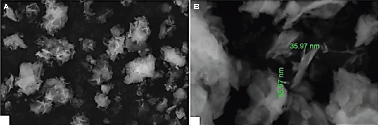
The morphology and dimensions of the synthesized NPs were examined using SEM.
Fig. 1 illustrates the successful synthesis of ZnO NPs. The ZnO NPs exhibited a non-spherical shape with consistent distribution. As indicated in
Fig. 1, the size of the produced particles varied between 35.9 nm and 111.1 nm.
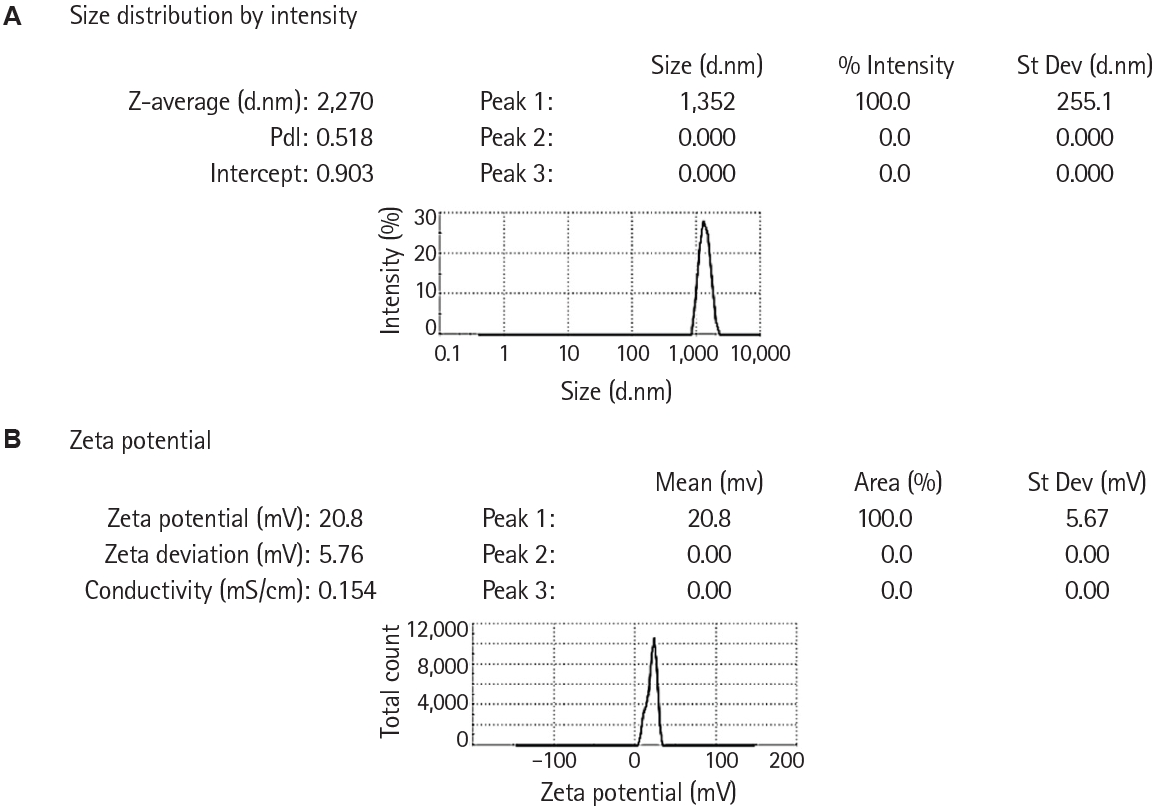
As shown in
Fig. 2, the main peak is at about 1,352 nm in diameter, meaning most particles are a little over 1 µm in size. The Z‑average size is 2,270 nm and the polydispersity index is 0.518, indicating a broad and somewhat heterogeneous size distribution rather than a very monodisperse sample. No secondary peaks are reported (Peak 2 and 3 are 0), so there is no clear evidence of separate smaller or larger particle populations, just 1 broad main population.
On the other hand, the zeta potential peak is at about +20.8 mV, with all of the area (100%) in this single peak. This value indicates a moderately positive surface charge: the particles have some electrostatic repulsion and colloidal stability, but they are not as strongly stabilized as systems with |ζ| above about 30 mV. The standard deviation of 5.76 mV shows a relatively tight distribution around this charge value, meaning most particles carry a similar surface charge.
S. mansoni adult worm burden
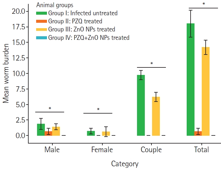
The administration of PZQ or ZnO NPs or the combination of both resulted in a substantial decrease in the overall worm burden, demonstrating statistical significance (
P<0.001) when compared to the infected control group that did not receive treatment (
Fig. 3).
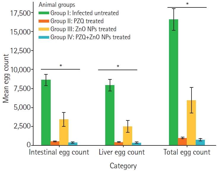
In relation to the egg counts of S. mansoni, the animals that received treatment exhibited a significant decrease in both hepatic and intestinal egg counts with (P<0.001) when compared to the infected non-treated control group.
As shown in
Fig. 4, animals that received a combination of both ZnO NPs and PZQ showed the highest reduction rate of total hepatic and intestinal egg counts (reduction of 95.4%) when compared to infected non-treated control group to those animals that received treatment. The group treated with PZQ demonstrated a substantial reduction in egg densities (reduction of 94.03%). This was followed by the animals treated with ZnO NPs, which showed a 63.9% reduction in total egg count.
As shown in
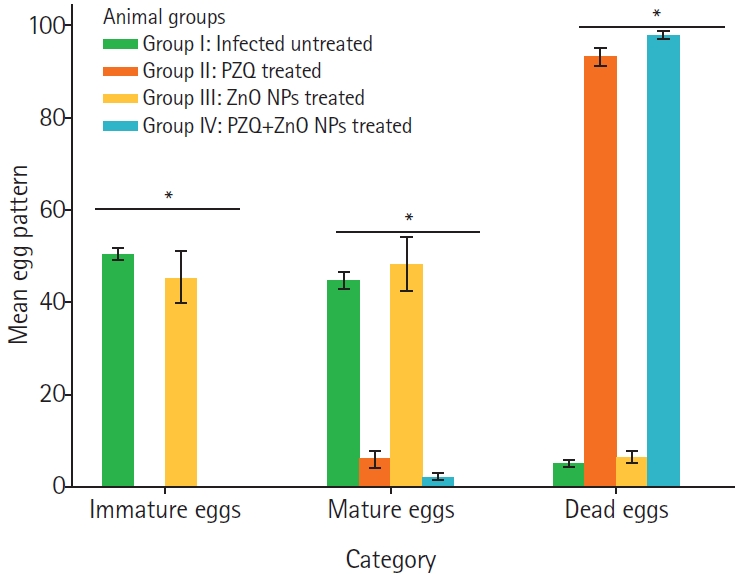
Fig. 5, the 3 treatment regimens significantly changed the oogram patterns when compared to the infected-untreated group. The highest percentage of dead eggs was recorded among ZnO NPs + PZQ-treated mice (97.8% versus 5% in infected untreated animals
P<0.002), as compared to infected-untreated animals (1.60%).
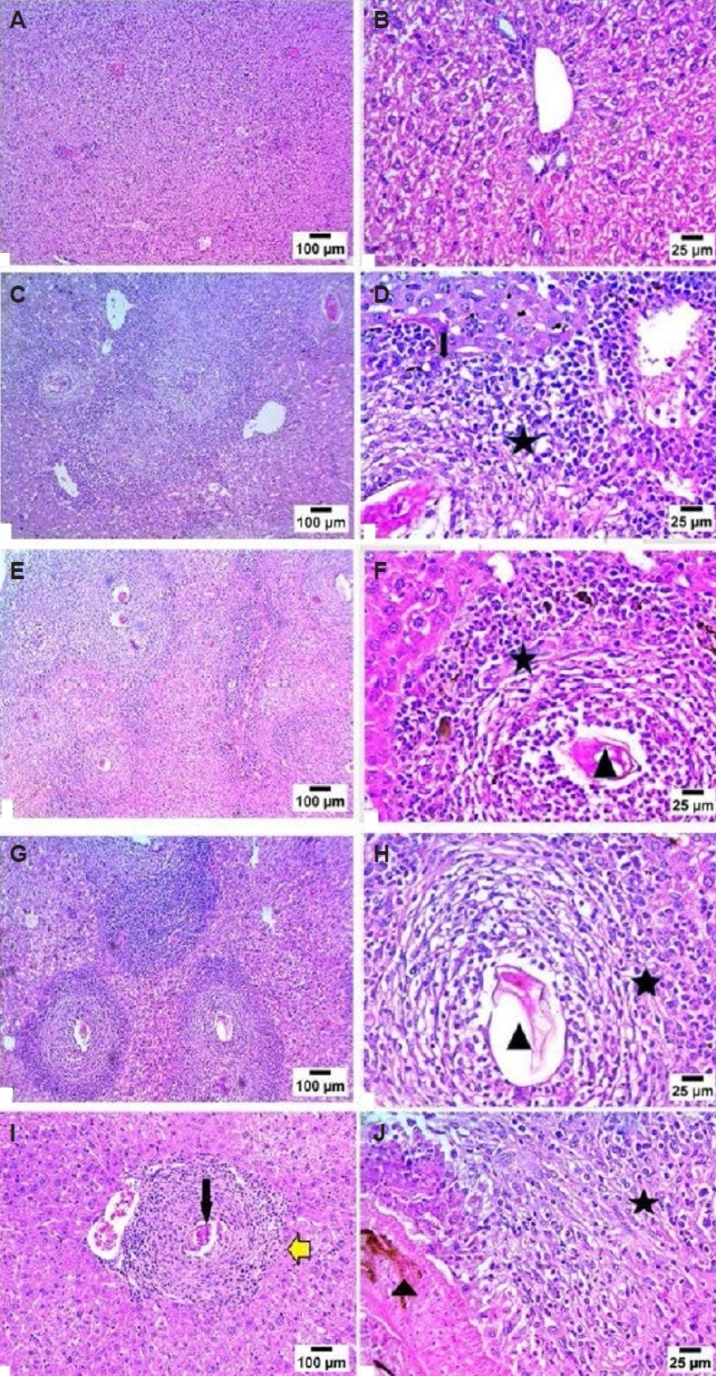
Microscopic analysis of liver sections from normal mice revealed the typical cellular structure of hepatic lobules. The normal arrangement of hepatic cords radiating from the central veins to the lobule’s periphery was evident, with narrow blood sinusoids lined by endothelial cells separating the cell cords. In contrast, the liver parenchyma of the infected untreated control group exhibited numerous chronic granulomatous lesions. These granulomas were primarily composed of parasite ova containing miracidia, surrounded by infiltrates of inflammatory cells. A notable feature of the granulomas was concentric fibrosis, characterized by numerous fibroblasts encircling the entrapped ova. Additionally, peri-granulomatous hepatocytes exhibited areas of necrosis and hydropic degeneration (
Fig. 6).
Histological examination of liver sections from both the PZQ and ZnO NPs-treated groups showed a significant improvement compared to the non-treated infected group. This improvement was evidenced by a reduction in both the number and size of granulomas (
Table 1). Moreover, there was a decrease in the number of bilharzial ova, the extent of hepatic fibrosis, and the infiltration of chronic inflammatory cells in these treated groups. Our results indicated a significant reduction in the average number of granulomas in both treated groups (
Table 1).
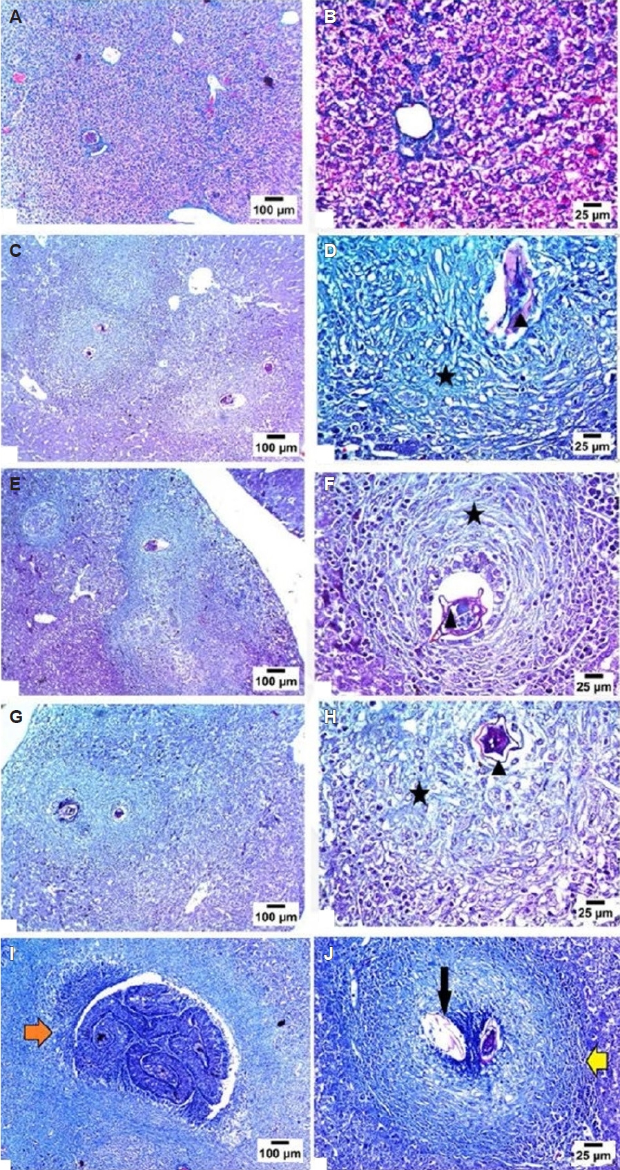
Liver fibrosis was evaluated utilizing Masson’s trichrome staining. In the sections from the negative control group, the central vein and portal tract walls displayed a thin layer of collagen fibers, signifying normal collagen deposition. Conversely, the infected non-treated group revealed a significant increase in collagen fibers surrounding granulomas, as well as periportal fibrosis and portal-portal fibrous bridging. In contrast, both the PZQ and ZnO NPs-treated groups demonstrated a decrease in collagen deposition around the granulomas and portal tracts when compared to the infected untreated mice (
Fig. 7).
Discussion
The present study demonstrates that green synthesized ZnO NPs exert significant anti schistosomal and hepato protective effects in a murine model of
S. mansoni infection, and that their combination with PZQ enhances therapeutic efficacy beyond PZQ monotherapy. The marked reduction in adult worm burden following treatment with PZQ, ZnO NPs, or their combination—each significantly lower than in infected untreated controls—confirms that both agents interfere with parasite survival. Of note, the ZnO NPs + PZQ regimen achieved nearly complete suppression of worm burden (99%), surpassing the already high efficacy of PZQ alone (96.28%), whereas ZnO NPs alone produced a smaller but still appreciable 21% reduction. This is consistent with previous research that has highlighted the anti-parasitic properties of various NPs, which are thought to induce oxidative stress and disrupt essential metabolic pathways in the parasites [
6].
The decrease in hepatic and intestinal egg counts further supports the therapeutic potential of ZnO NPs. The combined ZnO NPs and PZQ treatment resulted in the most significant reduction in total egg burden (95.4%), closely followed by PZQ monotherapy (94.03%), while ZnO NPs alone achieved a 63.9% reduction. Because schistosomiasis pathology is primarily driven by the host’s immune response to tissue trapped eggs, reducing egg deposition and viability is expected to attenuate granulomatous inflammation and subsequent fibrosis. Oogram analysis supports this idea: the ZnO NPs and PZQ group exhibited the highest rate of dead eggs (97.8% compared to 5% in infected untreated controls), and all treated groups showed a marked shift towards dead and degenerated eggs, which aligns with reduced egg viability or production.
Histopathological examination of liver tissue offers direct evidence of these protective effects. Infected untreated mice developed numerous chronic granulomas, characterized by schistosome eggs embedded in fibroblast rich concentric fibrosis, along with associated inflammatory infiltrates and zones of hepatocellular necrosis and hydropic degeneration. In contrast, livers from PZQ and ZnO NP treated animals demonstrated a notable improvement, with fewer and smaller granulomas, a reduced egg load, diminished inflammatory cell infiltration, and less extensive fibrosis. Quantitative evaluation confirmed a significant decrease in the mean number of granuloma in both treatment groups. Masson’s trichrome staining further revealed that collagen deposition surrounding granulomas and portal tracts was considerably lower in treated mice compared to infected controls, highlighting the ability of ZnO NPs, similar to PZQ, to mitigate the development of schistosomal liver fibrosis—a crucial factor in morbidity associated with chronic infection [
31,
32].
The use of ginger extract for green synthesis adds a biocompatibility and sustainability advantage [
14]. Ginger is rich in polyphenols, gingerols, alkaloids, and proteins that can act as reducing agents and surface capping ligands, facilitating the conversion of zinc ions to ZnO NPs and stabilizing the resulting NPs against aggregation. Such capping may also influence the biological activity of the NPs, potentially imparting antioxidants, anti-inflammatory, or antimicrobial properties that add to the intrinsic effects of ZnO. Although repeated washing and FTIR analysis did not show prominent peaks attributable to ginger derived organic moieties, trace phytochemical residues adsorbed on the NP surface cannot be entirely excluded and may have contributed to the observed anti-schistosomal and hepato-protective effects [
31,
33-
35].
Several limitations should be acknowledged. The absence of a ginger extract only control and a group treated with chemically synthesized (non green) ZnO NPs precludes a clear dissection of the relative contributions of the inorganic ZnO core versus ginger derived surface components to the therapeutic outcome. Future work should include these controls to distinguish NPs-specific effects from those attributable to plant metabolites. Additionally, the mechanistic interpretation of liver protection is currently limited to histopathology and fibrosis scoring; inclusion of serum liver enzymes (e.g., alanine transaminase, aspartate transaminase), oxidative stress markers, antioxidants, and inflammatory mediators would provide a more comprehensive picture of the underlying molecular pathways. Finally, the observed aggregation in aqueous media suggests that formulation, dispersion behavior under physiological conditions, pharmacokinetics, and long term toxicity should be systematically evaluated before considering clinical translation.
In conclusion, green synthesized ZnO NPs represent a promising adjunct to PZQ therapy for schistosomiasis, exhibiting significant worm reduction, egg suppression, and anti fibrotic effects in a murine S. mansoni model. The combination of ZnO NPs with PZQ appears superior to PZQ monotherapy in several key parameters, warranting further investigation to optimize dosing regimens, clarify the detailed mechanisms of action, and assess safety for potential therapeutic use.
Notes
-
Author contributions
Conceptualization: El-kady AM, Khodary M. Data curation: El-kady AM, Wakid MH. Formal analysis: El-kady AM, Fadel EF. Funding acquisition: Al-Megrin WA, Elshabrawy HA. Investigation: Mohamed K, Alhazmi A, Elshazly H, Sayed E, Elshabrawy HA, El Hassan SM. Methodology: Spears MW, Khodary M, El Hassan SM. Project administration: El-kady AM, Wakid MH. Resources: El-kady AM. Software: El-kady AM, Fadel EF, Khodary M. Supervision: El-kady AM, Wakid MH. Validation: Alhazmi A, Elshazly H, Sayed E, Elshabrawy HA. Visualization: Mohamed K, Alhazmi A, Al-Megrin WA, Elshazly H, Sayed E, Spears MW. Writing – original draft: El-kady AM. Writing – review & editing: El-kady AM, Wakid MH.
-
Conflict of interest
The authors have no conflicts of interest to declare.
-
Funding
This work was supported by Princess Nourah bint Abdulrahman University Researchers Supporting Project number (PNURSP2026R39) at Princess Nourah bint Abdulrahman University, Riyadh, Saudi Arabia.
Fig. 1.The morphology and dimensions of the synthesized zinc oxide (ZnO) nanoparticles (NPs). (A) Scanning electron microscopy morphology of ZnO NPs showing non spherical structures. (B) The size of ZnO NPs as measured by scanning electron microscopy ranging from 35.9 to 111.1 nm (×60,000).
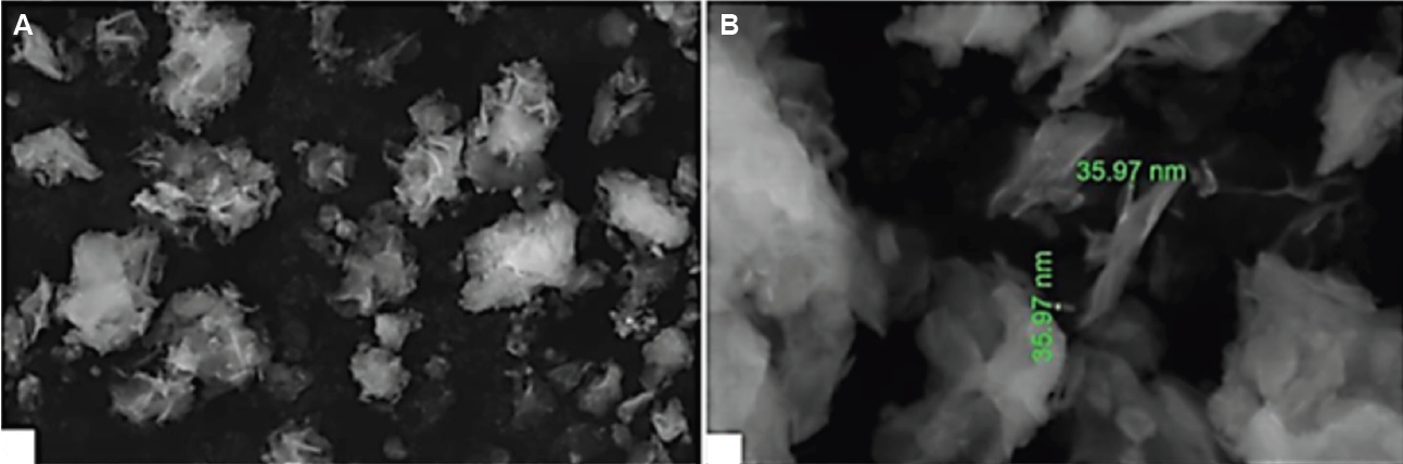
Fig. 2.Zeta potential characteristics of zinc oxide nanoparticles. (A) Size distribution by intensity. (B) Zeta potential as revealed by dynamic light scattering. The analysis revealed that the samples exhibited positive charges were measured 20.8±16.6 mV. The hydrodynamic diameters of the zinc oxide nanoparticles were determined to be 1,248 nm, with a polydispersity index (PDI) of 2,270.
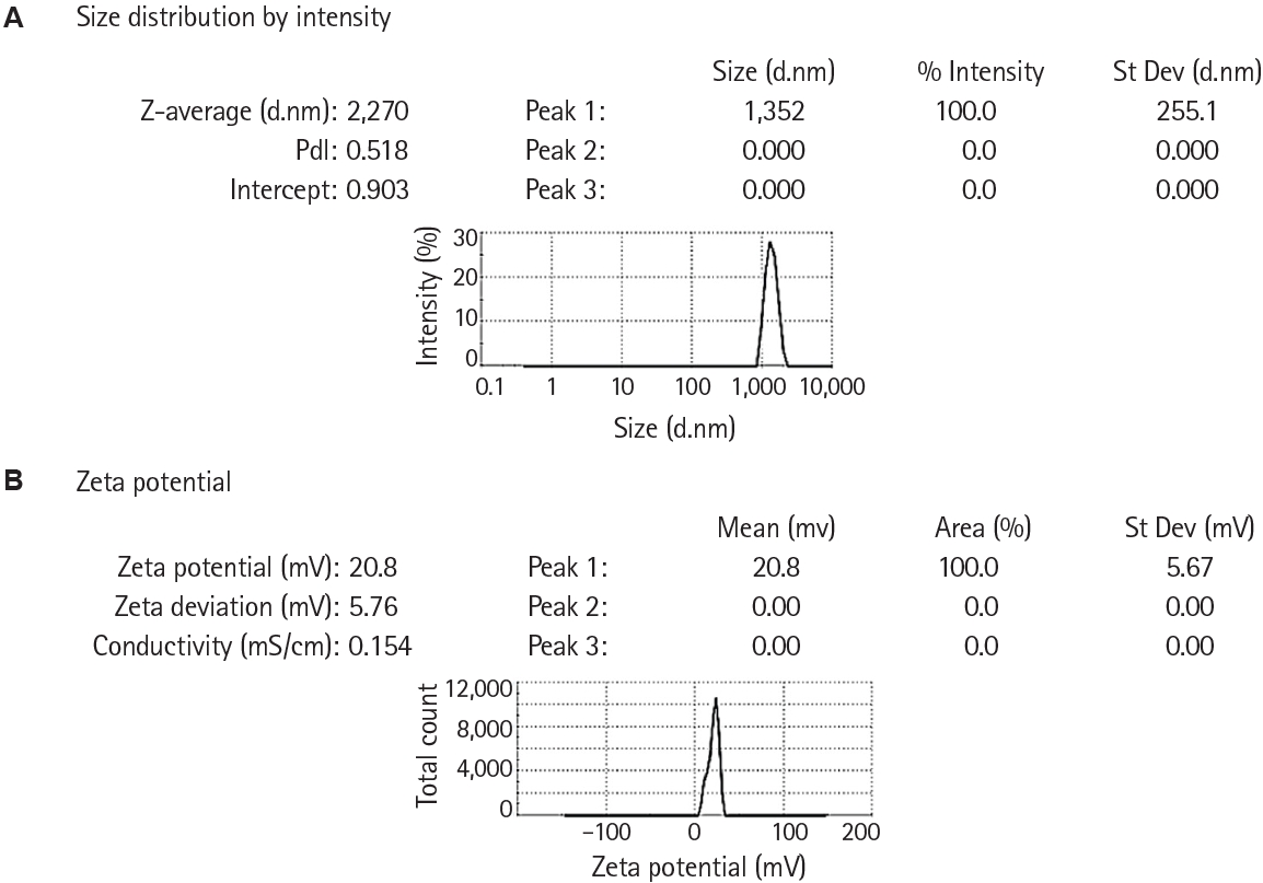
Fig. 3.
Schistosoma mansoni worm burden and total reduction percentage in each animal group. Data are expressed as means (n=10) and were analysed using one-way analysis of variance and Tukey as a post hoc test. * indicates significant difference between the infected untreated animal group on one hand and the treated groups on the other hand. PZQ, praziquantel; ZnO, zinc oxide; NPs, nanoparticles.
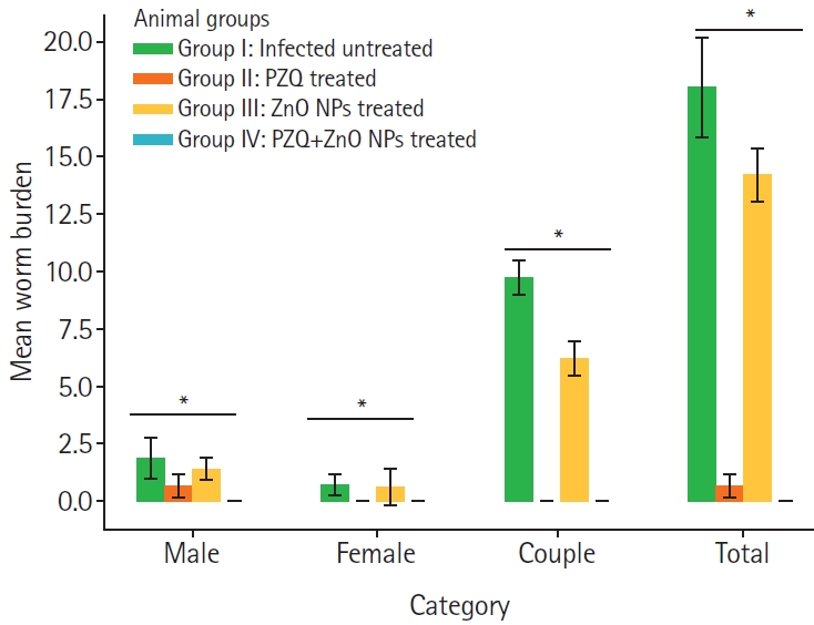
Fig. 4.Egg counts of Schistosoma mansoni in liver and intestinal tissues in each untreated and treated animal groups. Data are expressed as means (n=10) and were analysed using one-way analysis of variance and Tukey as a post hoc test. * indicates significant difference between the infected untreated animal group on one hand and the treated groups on the other hand. PZQ, praziquantel; ZnO, zinc oxide; NPs, nanoparticles.
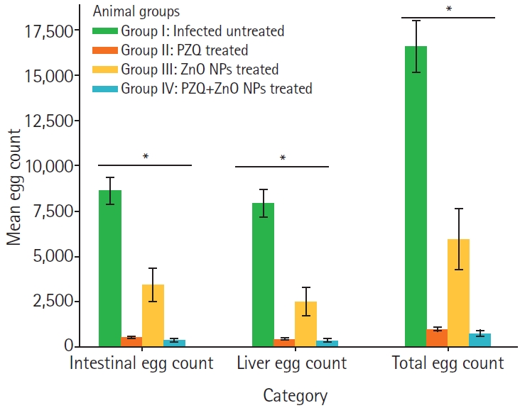
Fig. 5.Comparison of Schistosoma mansoni oogram patterns in treated and untreated animal groups. Data are expressed as means (n=10) and were analysed using one-way analysis of variance and Tukey as a post hoc test. * indicates significant difference between the infected untreated animal group on one hand and the treated groups on the other hand. PZQ, praziquantel; ZnO, zinc oxide; NPs, nanoparticles.
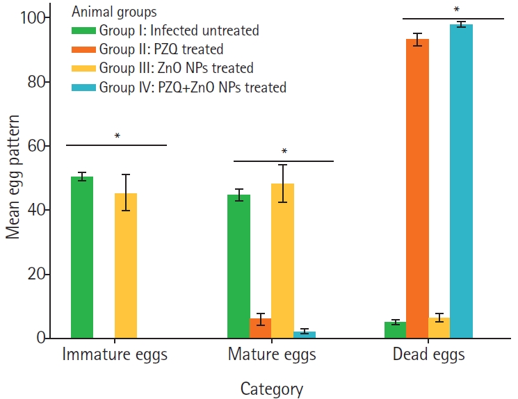
Fig. 6.Photomicrograph sections stained with hematoxylin-eosin showing the effect of various treatments on the Schistosoma induced hepatic granulomatous changes from each animal group investigated. (A, B) Sections from normal mice showing normal histological structure of the portal area. (C, D) Sections from S. mansoni infected untreated mice showing severe granulomatous changes in the portal triad, formed of fibrous connective tissue with inflammatory cells infiltration mainly eosinophils and lymphocytes (asterisk). (E, F) Sections from S. mansoni infected praziquantel treated mice with marked reduction of the number and diameter of S. mansoni egg granulomas in comparison with un-treated mice group. Liver sections showed granulomas in the portal triad, in which S. mansoni egg (arrowhead) is surrounded by fibrous connective tissue with inflammatory cells infiltration mainly eosinophils and lymphocytes (asterisk). (G, H) Sections from S. mansoni infected zinc oxide nanoparticles treated mice showing marked reduction of the number and diameter of S. mansoni egg granulomas in comparison with un-treated mice group. Sections showed granulomas change in the portal triad, in which S. mansoni (arrowhead) surrounded by fibrous connective tissue with inflammatory cells infiltration mainly eosinophils and lymphocytes (asterisk). (I, J) Sections from S. mansoni infected and treated with a combination of zinc oxide nanoparticle and praziquantel showing marked reduction of the number and diameter of S. mansoni egg granulomas in comparison with un-treated mice group. Sections showed granulomas change in the portal triad, in which adult S. mansoni (arrowhead) surrounded by fibrous connective tissue with inflammatory cells infiltration mainly eosinophils and lymphocytes (asterisk). (A, C, E, G, I) Magnification ×100, (B, D, F, H, J) Magnification ×400.
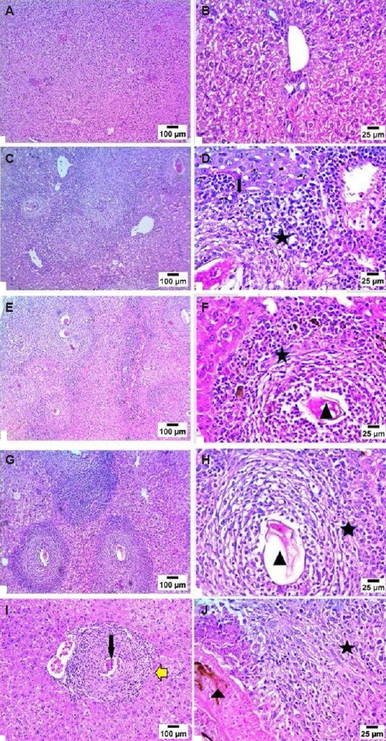
Fig. 7.Photomicrograph sections stained with Masson’s trichrome evaluating hepatic fibrosis from each animal group investigated. (A, B) Photomicrograph showing normal histological structure of portal area with no fibrosis formation. (C, D) Photomicrograph showing Schistosoma parasite (arrowhead) granuloma with marked infiltration of fibrous connective tissue (asterisk). (E, F) Photomicrograph showing Schistosoma parasite (arrowhead) granuloma with significant reduction of fibrous connective tissue infiltration (asterisk) in comparison to infected untreated mice. (G, H) Photomicrograph showing Schistosoma parasite (arrowhead) granuloma with significant reduction of fibrous connective tissue infiltration (asterisk) in comparison to infected untreated mice. (I, J) Photomicrograph showing Schistosoma parasite (orange arrow) granuloma with significant reduction of fibrous connective tissue infiltration in comparison to infected untreated mice. (I, J) Sections from S. mansoni infected and treated with a combination of zinc oxide nanoparticle and praziquantel showing fibrocellular granulomas (yellow arrow) and a dead-worm granuloma (orange arrow). (A, C, E, G, I) Magnification ×100, (B, D, F, H, J) Magnification ×400.
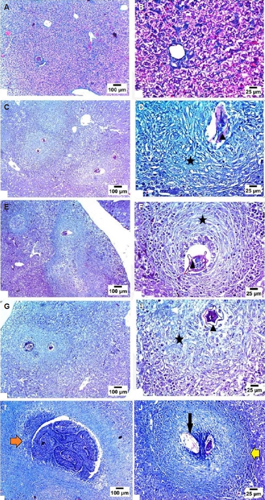
Table 1.Characteristics of the number and size of granulomas due to Schistosoma mansoni eggs in treated and untreated animal groups
Table 1.
|
Count |
Diameter |
Cellular granuloma |
Fibrocellular granuloma |
Intact eggs |
Degenerated eggs |
|
Animal group |
No. |
Reduction |
Size (µm) |
Reduction |
|
Group I. Infected untreated |
11.8 |
None |
294.3 |
None |
25 |
75 |
75 |
25 |
|
Group II. PZQ treated |
4.8 |
59.3 |
200 |
31.9 |
5 |
95 |
25 |
75 |
|
Group III. ZnO NPs treated |
9.7 |
17.8 |
261.7 |
11.1 |
40 |
60 |
75 |
25 |
|
Group IV. PZQ+ ZnO NPS treated |
6.5 |
45 |
258.3 |
12.4 |
5 |
95 |
25 |
75 |
References
- 1. LoVerde PT. Schistosomiasis. In: Toledo R, Fried B, editors. Digenetic trematodes. Springer; 2024. p. 75-106.
- 2. Song LJ, Yin XR, Mu SS, et al. The differential and dynamic progression of hepatic inflammation and immune responses during liver fibrosis induced by Schistosoma japonicum or carbon tetrachloride in mice. Front Immunol 2020;11:570524. https://doi.org/10.3389/fimmu.2020.570524
- 3. Doenhoff MJ, Cioli D, Utzinger J. Praziquantel: mechanisms of action, resistance and new derivatives for schistosomiasis. Curr Opin Infect Dis 2008;21:659-67. https://doi.org/10.1097/QCO.0b013e328318978f
- 4. Melman SD, Steinauer ML, Cunningham C, et al. Reduced susceptibility to praziquantel among naturally occurring Kenyan isolates of Schistosoma mansoni. PLoS Negl Trop Dis 2009;3:e504. https://doi.org/10.1371/journal.pntd.0000504
- 5. Kaittanis C, Santra S, Perez JM. Emerging nanotechnology-based strategies for the identification of microbial pathogenesis. Adv Drug Deliv Rev 2010;62:408-23. https://doi.org/10.1016/j.addr.2009.11.013
- 6. Dkhil MA, Al-Quraishy S, Wahab R. Anticoccidial and antioxidant activities of zinc oxide nanoparticles on Eimeria papillata-induced infection in the jejunum. Int J Nanomedicine 2015;10:1961-8. https://doi.org/10.2147/IJN.S79944
- 7. Devi HS, Boda MA, Shah MA, Parveen S, Wani AH. Green synthesis of iron oxide nanoparticles using Platanus orientalis leaf extract for antifungal activity. Green Process Synth 2019;8:38-45. https://doi.org/10.1515/gps-2017-0145
- 8. Alsammarraie FK, Wang W, Zhou P, Mustapha A, Lin M. Green synthesis of silver nanoparticles using turmeric extracts and investigation of their antibacterial activities. Colloids Surf B Biointerfaces 2018;171:398-405. https://doi.org/10.1016/j.colsurfb.2018.07.059
- 9. Kataria N, Garg VK. Green synthesis of Fe3O4 nanoparticles loaded sawdust carbon for cadmium (II) removal from water: regeneration and mechanism. Chemosphere 2018;208:818-28. https://doi.org/10.1016/j.chemosphere.2018.06.022
- 10. Nasrollahzadeh M, Mohammad Sajadi S. Pd nanoparticles synthesized in situ with the use of Euphorbia granulate leaf extract: catalytic properties of the resulting particles. J Colloid Interface Sci 2016;462:243-51. https://doi.org/10.1016/j.jcis.2015.09.065
- 11. Chitra K, Annadurai G. Antimicrobial activity of wet chemically engineered spherical shaped ZnO nanoparticles on food borne pathogen. Int Food Res J 2013;20:59-64.
- 12. Dwivedi S, Wahab R, Khan F, et al. Reactive oxygen species mediated bacterial biofilm inhibition via zinc oxide nanoparticles and their statistical determination. PLoS One 2014;9:e111289. https://doi.org/10.1371/journal.pone.0111289
- 13. Emamifar A, Kadivar M, Shahedi M, Soleimanian-Zad S. Evaluation of nanocomposite packaging containing Ag and ZnO on shelf life of fresh orange juice. Innov Food Sci Emerg Technol 2010;11:742-8. https://doi.org/10.1016/j.ifset.2010.06.003
- 14. Król G, Fortunka K, Majchrzak M, et al. Metallic nanoparticles and core-shell nanosystems in the treatment, diagnosis, and prevention of parasitic diseases. Pathogens 2023;12:838. https://doi.org/10.3390/pathogens12060838
- 15. Ranjan AP, Mukerjee A, Helson L, Vishwanatha JK. Scale up, optimization and stability analysis of Curcumin C3 complex-loaded nanoparticles for cancer therapy. J Nanobiotechnology 2012;10:38. https://doi.org/10.1186/1477-3155-10-38
- 16. Abd El Wahab WM, El-Badry AA, Mahmoud SS, et al. Ginger (Zingiber Officinale)-derived nanoparticles in Schistosoma mansoni infected mice: hepatoprotective and enhancer of etiological treatment. PLoS Negl Trop Dis 2021;15:e0009423. https://doi.org/10.1371/journal.pntd.0009423
- 17. Muchirah PN, Yole D, Kutima H, et al. Determination of effective praziquantel dose in different mouse strains: BALB/c and Swiss mice in treatment of Schistosoma mansoni. J Clin Immunol Immunopathol Res 2012;4:12-21. https://doi.org/10.5897/JCIIR12.004
- 18. Melk MM, El-Hawary SS, Melek FR, et al. Antiviral activity of zinc oxide nanoparticles mediated by Plumbago indica L. extract against herpes simplex virus type 1 (HSV-1). Int J Nanomedicine 2021;16:8221-33. https://doi.org/10.2147/IJN.S339404
- 19. Rescignano N, Amelia M, Credi A, Kenny JM, Armentano I. Morphological and thermal behavior of porous biopolymeric nanoparticles. Eur Polym J 2012;48:1152-9. https://doi.org/10.1016/j.eurpolymj.2012.05.005
- 20. Cohen-Sela E, Chorny M, Koroukhov N, Danenberg HD, Golomb G. A new double emulsion solvent diffusion technique for encapsulating hydrophilic molecules in PLGA nanoparticles. J Control Release 2009;133:90-5. https://doi.org/10.1016/j.jconrel.2008.09.073
- 21. Al-Refai MF, Wakid MH. Prevalence of intestinal parasites and comparison of detection techniques for soil-transmitted helminths among newly arrived expatriate labors in Jeddah, Saudi Arabia. PeerJ 2024;12:e16820. https://doi.org/10.7717/peerj.16820
- 22. Bahwaireth EO, Wakid MH. Molecular, microscopic, and immunochromatographic detection of enteroparasitic infections in hemodialysis patients and related risk factors. Foodborne Pathog Dis 2022;19:830-8. https://doi.org/10.1089/fpd.2022.0024
- 23. Campbell WC. Trichinella and trichinosis. Springer Science & Business Media; 2012.
- 24. El-kady AM, Hassan AS, Mohamed K, et al. Zinc oxide nanoparticles produced by Zingiber officinale ameliorates acute toxoplasmosis-induced pathological and biochemical alterations and reduced parasite burden in mice model. PLoS Negl Trop Dis 2023;17:e0011447. https://doi.org/10.1371/journal.pntd.0011447
- 25. Duvall RH, DeWitt WB. An improved perfusion technique for recovering adult schistosomes from laboratory animals. Am J Trop Med Hyg 1967;16:483-6. https://doi.org/10.4269/ajtmh.1967.16.483
- 26. Kamel IA, Cheever AW, Elwi AM, Mosimann JE, Danner R. Schistosoma mansoni and S. haematobium infections in Egypt. I. Evaluation of techniques for recovery of worms and eggs at necropsy. Am J Trop Med Hyg 1977;26:696-701. https://doi.org/10.4269/ajtmh.1977.26.696
- 27. Muema JM, Obonyo MA, Njeru SN, Mwatha JK. Antischistosomal effects of selected methanolic plant extracts in Swiss Albino mice infected with Schistosoma mansoni. Eur J Med Plants 2015;9:1-11. https://doi.org/10.9734/EJMP/2015/16953
- 28. Jatsa HB, Kenfack CM, Simo DN, et al. Schistosomicidal, hepatoprotective and antioxidant activities of the methanolic fraction from Clerodendrum umbellatum Poir leaves aqueous extract in Schistosoma mansoni infection in mice. BMC Complement Altern Med 2015;15:248. https://doi.org/10.1186/s12906-015-0788-z
- 29. Pellegrino J, Oliveira CA, Faria J, Cunha AS. New approach to the screening of drugs in experimental schistosomiasis mansoni in mice. Am J Trop Med Hyg 1962;11:201-15. https://doi.org/10.4269/ajtmh.1962.11.201
- 30. El-Kott AF, Mohammed RT, Ismail NR. Efficacy of garlic and mirazid in treatment of the liver granuloma in mice infected with Schistosoma mansoni. Res J Parasitol 2011;6:151-9. https://doi.org/10.3923/jp.2011.151.159
- 31. Ali EM, Abdalameer NK, Khalaph KA, et al. Green reduction approach for the synthesis of copper oxide nanoparticles using ginger extract and evaluation of their free radical scavenging activity. Eur Phys J Plus 2025;140:928. https://doi.org/10.1140/epjp/s13360-025-06873-1
- 32. Ali SB, Mohamed AS, Fahmy SR, Eet al. Anthelmintic and hepatoprotective activities of the green-synthesized zinc oxide nanoparticles against Parascaris equorum infection in rats. Acta Parasitol 2024;69:283-301. https://doi.org/10.1007/s11686-023-00728-4
- 33. Abd Jabar NA, Nor-Khaizura MAR, Ismail SI, et al. Are Bentong ginger (Zingiber officinale) biosynthesized silver nanoparticles safe and effective? An optimization, characterization, and toxicity evaluation study. OpenNano 2025;21:100224. https://doi.org/10.1016/j.onano.2024.100224
- 34. Cherik IT, Divsalar A, Angaji SA, et al. Green synthesis and characterization of ginger-extract-based oxali-palladium nanoparticles for colorectal cancer: downregulation of REG4 and apoptosis induction. Nanotechnol Rev 2023;12:20230154. https://doi.org/10.1515/ntrev-2023-0154
- 35. Mehrotra S, Goyal V, Dimkpa CO, Chhokar V. Green synthesis and characterization of ginger-derived silver nanoparticles and evaluation of their antioxidant, antibacterial, and anticancer activities. Plants (Basel) 2024;13:1255. https://doi.org/10.3390/plants13091255


 , Majed H. Wakid2,3,*