Abstract
Blastocystis is a genus of unicellular heterokont parasites belonging to a group of organisms known as Stramenopiles, which includes algae, diatoms, and water molds. Blastocystis includes several species that habitat in the gastrointestinal tracts of organisms as diverse as humans, farm animals, birds, rodents, reptiles, amphibians, fish, and cockroaches. It is important to public health and distributed globally, but its prevalence in dogs in Korea has not been reported to date. Here, we collected 787 canine fecal samples and assessed Blastocystis infection by age, sex, region, season, and diarrhea symptoms. We determined Blastocystis subtypes using phylogenetic analyses based on 18S rRNA gene sequences. We identified, 10 Blastocystis positive samples (1.3%). A higher proportion of infected dogs was asymptomatic; however, infection rates did not significantly differ according to region, age, sex, and season. Phylogenetic analysis showed that the Blastocystis sp. identified belonged to 4 subtypes (STs), ST1, ST5, ST10, and ST14, thus revealed the genetic diversity of Blastocystis sp. in dogs Korean. This is first report on the presence of Blastocystis sp. in dogs Korean. This study revealed a lower infection rate than expected and differed from previous studies in STs. Further studies are warranted to observe the national infection status of Blastocystis in dogs and the genetic characteristics of this genus.
-
Key words: Blastocystis, phylogeny, dog, prevalence, subtyping, 18S rRNA
Recently, the Korean pet market has reached 2 trillion ton, and approximately 20% of households own pets. These pets are a potential source of infection for their owners. A variety of pathogens can induce zoonotic diseases, among which, the intestinal protozoan,
Blastocystis, has never been studied in Korean dogs.
Blastocystis is a genus of single-celled heterokont parasites belonging to Stramenopiles including algae, diatoms, and water molds.
Blastocystis inhabit in the gastrointestinal tracts of host species [
1]. As
Blastocystis has low host specificity, humans as well as several animals, can be easily infected and facilitate its transmission. The most representative species found in humans is
Blastocystis hominis with subtypes (STs) belonging to ST 1–10 [
2]. Furthermore, many other STs are found in various hosts. Recently, 4 species of
Blastocystis were reported from nonhuman hosts:
B. galli from chickens [
3],
B. anatis from domestic ducks [
4],
B. anseri from domestic geese [
5], and
B. lapemi from a sea snake [
6].
Blastocystis is globally distributed, with a higher prevalence in developing countries. As these are intestinal protozoans, some patients present with gastrointestinal symptoms such as diarrhea, whereas others experienced no symptoms. The pathogenicity of
Blastocystis remains controversial. The infection spreads via water or food contaminated with the feces of infected hosts, and effective therapies have not been determined to date. The prevalence of
Blastocystis in dogs has never been examined in Korea. The present study investigated the prevalence of
Blastocystis sp., in Korean dogs using molecular method, and compared the phylogenetic characteristics of these species with those reported in other animals.
The statistical sample size was determined using a formula with an expected disease prevalence of 20%, a confidence level of 95%, and an accepted absolute error of 5%, with a simple random sampling strategy [
7]. A minimum of 246 samples were required based on this formula. We collected stool samples from 787 dogs randomly selected from shelter homes, animal hospitals, and Korean military establishments spread across Korea during 2016–2020. Ethical approval was not needed as fecal sample collection did not harm the animals. The samples were sent to the Laboratory of Veterinary Parasitology at Kyungpook National University College of Veterinary Medicine (Daegu, Korea) for parasite examination. Basic information about each dog was collected, in terms of its breeding environment, age, sex, breed, and the presence of diarrhea. Based on the breeding environment (shelter dogs, companion dogs, others including military dogs, police dogs, and special purpose dogs), there were 317 shelter dogs, 337 companion dogs, and 133 other dogs. By age, the animals were classified into 5 groups: puppy (less than 3 months old), junior (4–7 months), adult (8 months to 7 years), and senior (older than 8 years). There were 33 puppies, 25 juniors, 365 adults, 135 seniors, and 229 dogs of unknown age. There were 338 males, 298 females, and 151 dogs of unknown sex. The number of samples by region was as follows: 341 from northern, 200 from central, 226 from southern, and 20 from unknown regions of Korea. A total of 211 samples were collected in spring, 213 in summer, 248 in fall, and 115 in winter. Diarrheal symptoms were present in 100 dogs, while 560 had no diarrhea symptoms. The symptoms of the remaining 127 dogs were unknown.
Genomic DNA was extracted from fecal samples using the QIAamp Fast DNA Stool Mini Kit (Qiagen, Hilden, Germany). Each DNA sample was subjected to a PCR assay using a Bioneer HotStart PCR premix kit (Bioneer, Daejeon, Korea). For detecting
Blastocystis, nested PCR was performed that amplified a 480 bp region of the 18S rRNA. The primer sequences used were as follows: forward: (5′-GGAGGTAGTGACAATAAATC-3′) and reverse: (5′-TGCTTTCGCACTTGTTCATC-3′) as recently reported [
8,
9]. These sequences targeted the conserved regions of the 18S rRNA sequences of
Blastocystis available in GenBank. PCR-positive samples were sent to Macrogen (Daejeon, Korea) for sequencing.
All sequences were subjected to BLAST searches against the GenBank database to confirm the specimens as Blastocystis sp. Statistical analyses were performed with the χ2 test using SPSS V.21.0 and results with P<0.05 were considered statistically significant. Phylogenetic analyses were performed using MEGA 6.0. Phylogenetic inferences were made using the maximum likelihood method with 1,000 bootstrap replicates.
Out of 787 dog fecal samples, 10 feces (1.3%) were positive for
Blastocystis sp. (
Table 1), which included 1 shelter dog (0.1%), 6 companion dogs (0.8%), and 3 other dogs (0.4%). We also assessed the
Blastocystis prevalence according to age, sex, diarrhea status, region, and season. Based on age,
Blastocystis sp. was found in 2.2% (8/365) adults and 1.5% (2/135) seniors. According to sex, 0.9% (3/338) males, 2.0% (6/298) females, and 0.7% (1/151) sex–unknown dogs were positive for
Blastocystis sp. Among 560 dogs without diarrheal symptoms, 10 samples tested positive for
Blastocystis. By region, 1.8% (6/341), 1.0% (2/200), and 0.9% (2/226) samples were positive for
Blastocystis sp. from the northern, central, and southern regions, respectively. By season, 0.5% (1/211), 0.5% (1/213), 2.8% (7/248), and 0.95 (1/115) samples collected in the spring, summer, fall, and winter, respectively, tested positive for
Blastocystis sp. No significant differences were found in the risk factors of age, sex, diarrhea, region, and season. Although female dogs from the northern region during fall season demonstrated a higher positive rate than those in the other groups, but the difference was not statistically significant.
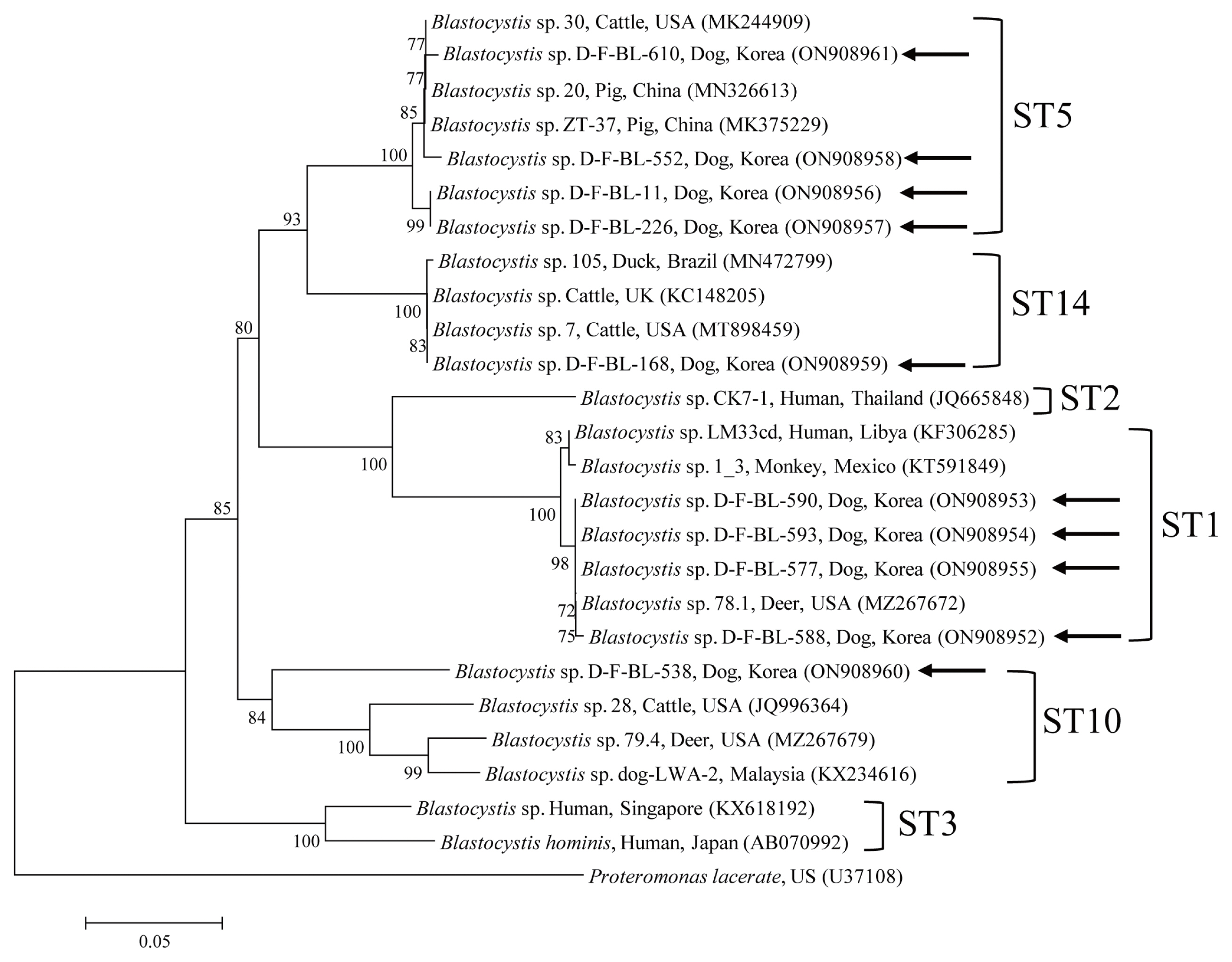
A phylogenetic analysis of all 18S rRNA nucleotide sequences of
Blastocystis sp. obtained in this study was performed against reference sequences retrieved from GenBank (
Fig. 1). According to the sequencing results, the 10 sequences were divided into 4 STs: ST1, ST5, ST10, and ST14. The ST distribution showed that the dominant types were ST1 (4/10, 40%) and ST5 (4/10, 40%), followed by ST10 (1/10, 10%) and ST14 (1/10, 10%). The 4 sequences of ST1 and ST5 found in the present study shared 99.8–100% and 97.5–98.6% identity, respectively, with each other. They also had 98.9–100% and 97.5–99.3% identity with the 18S rRNA sequences of previously reported
Blastocystis sp. isolates, respectively. The ST10 and ST14 sequences had 95.6–97.8% and 99.8–100% identity with the 18S rRNA sequences of previously reported
Blastocystis species. The 10 sequences reported in this study were registered in the GenBank, with the following accession numbers: 4 sequences belonging to ST1 (ON908952-ON908955), 4 sequences belonging to ST5 (ON908956-ON908958, ON908961), 1 sequence belonging to ST10 (ON908960), and 1 sequence belonging to ST14 (ON908959), respectively.
The prevalence of
Blastocystis infection varies according to animal groups, countries, and STs. In addition, it can be affected by diagnostic tools and status of hygiene.
Blastocystis infections in southeast Asian countries have been reported in various animals, such as companion animals, poultry, non-human primates, and livestock, over the last decade [
10]. The prevalence of
Blastocystis infection has also been examined in several countries around the world [
11]. The infection rate of
Blastocystis in dogs was found to be 2.5% (2/80), 1.3% (1/80), and 24% (19/80) in Australia, Cambodia, and India [
12]. In Korea,
Blastocystis infection has been detected in pigs [
13], cats [
14], wild boars [
15], Korean water deer [
16], and cattle [
17], while none has evaluated
Blastocystis infection in dogs. We, for the first time, found a low prevalence of
Blastocystis sp. in Korea.
In general, a high infection rate is expected in shelter dogs, but we found that only 1 shelter dog (0.1%) was infected. In contrast, 6 dogs (0.8%) from the companion dog group were infected, indicating a higher infection rate among this group. Companion dogs are kept outside or inside the home. Outside the home, dogs may ingest the feces of an infected animal while walking or come in contact with other infected dogs; inside the home, the possibility of animals being infected by humans should be considered due to the low host specificity of Blastocystis. Furthermore, humans should be careful considering the high rate of Blastocystis infection in companion dogs. The three infected dogs (0.4%) were detected in the other groups, such as military dogs, police dogs, and special purpose dogs. Since they live in various special environments, they might be highly susceptible to infection.
In contrast to general findings, we recognized that the infection rate was higher in adult dogs than in younger or older dogs. Typically, hosts with low immunity have a higher rate of protozoan infection and show more severe symptoms. In humans, a higher prevalence of protozoal disease has been reported in adults than in children; young adults appear to have the highest rates of infection and infection rates may decrease in older adults [
18]. However, a recent study observed a statistically significant reduction in infection rates in older children than younger children [
19]. Our result indicates that
Blastocystis sp. shows higher infectivity in adult dogs in Korea. However, the number of samples from young dogs was small. Moreover, in Korea, the coverage rate and preventative anti-amebic dosage rate is high in young dogs, which may have influenced the results.
Significantly, all positive samples were from dogs without diarrhea. However, as diarrhea is not the only symptom of Blastocystis infection, it is necessary to compare infection rates with other symptoms. Furthermore, there may be a reason for the lower number of diarrhea cases than that of asymptomatic cases. There may also be several causes for diarrhea other than Blastocystis; therefore, these causes of diarrhea symptoms should be ruled out to obtain more accurate results.
In other countries, STs such as ST1–8, and ST10 show large differences across regions [
10]. The stray dogs in India carry a diverse range of
Blastocystis STs including ST1, 4, 5, and 6. The ST2 was detected in one dog in Cambodia, and ST1 was detected in 2 dogs in Australia [
11]. In this study, phylogenetic analysis confirmed the presence of 4 genetic
Blastocystis STs in Korean dogs. The ST1 and ST5 strains were predominant in this study and were closely related to species isolated from humans, indicating the zoonotic potential of this genus. ST1 and ST5 are widely distributed throughout Korea, whereas ST10 and ST14 were only detected in the central and northern region, respectively. Among them, 3 dogs with ST1 were raised in the same military unit. They may have been cross-infected with Blastocystis sp. in the same environment.
To the best of our knowledge, this is first report of Blastocystis infection in dogs in Korea. Our finding differs from those of previous studies in terms of an overall low infection rate. Our result indicates a low rate of Blastocystis infection rate in Korean dogs, despite the global distribution of these organisms. However, our results should be verified in future studies. In addition, care should be taken to avoid cross-infection considering the zoonotic potential of Blastocystis sp. in humans.
In conclusion, of 787 dog fecal samples examined in this study, 10 (1.3%) tested positive for Blastocystis sp. Phylogenetic analysis showed that the Blastocystis sp. identified in dogs belonged to ST1, ST5, ST10, and ST14. Of these, ST1 and ST5 have zoonotic potential. Phylogenetic analysis revealed genetic diversity of Blastocystis sp. in dogs across Korea. The infection rate was unexpectedly low, and there were no symptoms of diarrhea in the infected dogs. The prevalence of Blastocystis in the dogs in Korea has not been reported previously, and genotyping of the remaining Blastocystis isolates is ongoing. Future studies should thus include additional sequences of more samples, relationship between infection and symptoms, and perform detailed phylogenetic analysis, genotyping, and survey across various animals.
Notes
-
The authors declare no conflicts of interest related to the publication of this study.
Fig. 1Phylogenetic tree of Blastocystis. A phylogenetic tree was constructed from the 18S rRNA sequences generated in this study using the maximum likelihood method based on the Tamura-Nei model (1,000 replicates). The sequences identified in this study are indicated by arrows.

Table 1Prevalence of Blastocystis based on the 18S rRNA gene in dog fecal samples collected in Korea during 2016–2020
Table 1
|
Group |
No. tested |
Shelter dog |
Companion dog |
Other |
Total |
|
Age (yr) |
|
Puppy (≤3 m) |
33 |
0 |
0 |
0 |
0 |
|
Junior (4–7 m) |
25 |
0 |
0 |
0 |
0 |
|
Adult (8 m–7 yr) |
365 |
1 (0.3) |
4 (1.1) |
3 (0.8) |
8 (2.2) |
|
Senior (≥8 yr) |
135 |
0 |
2 (1.5) |
0 |
2 (1.5) |
|
Unknown |
229 |
0 |
0 |
0 |
0 |
|
|
Sex |
|
Male |
338 |
0 |
0 (0.0) |
3 (0.9) |
3 (0.9) |
|
Female |
298 |
1 (0.3) |
5 (1.7) |
0 |
6 (2.0) |
|
Unknown |
151 |
0 |
1 (0.7) |
0 |
1 (0.7) |
|
|
Diarrhea |
|
Yes |
100 |
0 |
0 |
0 |
0 |
|
No |
560 |
1 (0.2) |
6 (1.1) |
3 (0.5) |
10 (1.8) |
|
Unknown |
127 |
0 |
0 |
0 |
0 |
|
|
Region |
|
Northern |
341 |
0 |
4 (1.2) |
2 (0.6) |
6 (1.8) |
|
Central |
200 |
0 |
2 (1.0) |
0 |
2 (1.0) |
|
Southern |
226 |
1 (0.4) |
0 (0.0) |
1 (0.4) |
2 (0.9) |
|
Unknown |
20 |
0 |
0 |
0 |
0 |
|
|
Season |
|
Spring |
211 |
0 |
1 (0.5) |
0 |
1 (0.5) |
|
Summer |
213 |
1 (0.5) |
0 |
0 |
1 (0.5) |
|
Fall |
248 |
0 |
4 (1.6) |
3 (1.2) |
7 (2.8) |
|
Winter |
115 |
0 |
1 (0.9) |
0 |
1 (0.9) |
References
- 1. Yoshikawa H, Wu Z, Howe J, Hashimoto T, Geok-Choo N, Tan KS. Ultrastructural and phylogenetic studies on Blastocystis isolates from cockroaches. J Eukaryot Microbiol 2007;54:33-37. https://doi.org/10.1111/j.1550-7408.2006.00141.x
- 2. Noël C, Dufernez F, Gerbod D, Edgcomb VP, Delgado-Viscogliosi P, Ho LC, Singh M, Wintjens R, Sogin ML, Capron M, Pierce R, Zenner L, Viscogliosi E. Molecular phylogenies of Blastocystis isolates from different hosts: implications for genetic diversity, identification of species, and zoonosis. J Clin Microbiol 2005;43:348-355. https://doi.org/10.1128/JCM.43.1.348-355.2005
- 3. Belova LM, Kostenko LA. Blastocystis galli sp. n. (Protista: Rhizopoda) from the intestine of domestic hens. Parazitologiia 1990;24:164-168. (In Russian).
- 4. Belova LM. Blastocystis anatis sp. n. (Rhizopoda, Lobosea) from Anas platyrhynchos. Zool Zh 1991;70:5-10. (In Russian).
- 5. Belova LM. Blastocystis anseri (Protista, Rhizopoda) from domestic goose. Parazitologiia 1992;26:80-82. (In Russian).
- 6. Teow WL, Ng GC, Chan PP, Chan YC, Yap EH, Zaman V, Singh M. A survey of Blastocystis in reptiles. Parasitol Res 1992;78:453-455. https://doi.org/10.1007/BF00931705
- 7. Thrusfield M. Veterinary Epidemiology. 3rd ed. Blackwell Publishing; Oxford, UK. 2005. p. 232-233.
- 8. Böhm-Gloning B, Knobloch J, Walderich B. Five subgroups of Blastocystis hominis from symptomatic and asymptomatic patients revealed by restriction site analysis of PCR-amplified 16S-like rDNA. Trop Med Int Health 1997;2:771-778. https://doi.org/10.1046/j.1365-3156.1997.d01-383.x
- 9. Santín M, Gómez-Muñoz MT, Solano-Aguilar G, Fayer R. Development of a new PCR protocol to detect and subtype Blastocystis spp. from humans and animals. Parasitol Res 2011;109:205-212. https://doi.org/10.1007/s00436-010-2244-9
- 10. Rauff-Adedotun AA, Mohd Zain SN, Farah Haziqah MT. Current status of Blastocystis sp. in animals from Southeast Asia: a review. Parasitol Res 2020;119:3559-3570. https://doi.org/10.1007/s00436-020-06828-8
- 11. Kaczmarek A, Rocka A, Wesołowska M, Gołąb E, Sałamatin R. Blastocystis isolates from a dog and their owners presenting with chronic diarrhoea. Dogs as reservoirs of Blastocystis: research in Poland and worldwide. Ann Parasitol 2020;66:573-579. https://doi.org/10.17420/ap6604.300
- 12. Wang W, Cuttell L, Bielefeldt-Ohmann H, Inpankaew T, Owen H, Traub RJ. Diversity of Blastocystis subtypes in dogs in different geographical settings. Parasit Vectors 2013;6:215. https://doi.org/10.1186/1756-3305-6-215
- 13. Paik S, Jung BY, Lee H, Hwang MH, Han JE, Rhee MH, Kim TH, Kwon OD, Kwak D. Molecular detection and subtyping of Blastocystis in Korean pigs. Korean J Parasitol 2019;57:525-529. https://doi.org/10.3347/kjp.2019.57.5.525
- 14. Kwak D, Seo MG. Genetic analysis of zoonotic gastrointestinal protozoa and microsporidia in shelter cats in South Korea. Pathogens 2020;9:894. https://doi.org/10.3390/pathogens9110894
- 15. Lee H, Seo MG, Oem JK, Kim YS, Lee SY, Kim J, Jeong H, Jheong WH, Kim Y, Lee WJ, Kwon OD, Kwak D. Molecular detection and subtyping of Blastocystis detected in wild boars (Sus scrofa) in South Korea. J Wildl Dis 2020;56:662-666. https://doi.org/10.7589/2019-04-092
- 16. Kim KT, Noh G, Lee H, Kim SH, Jeong H, Kim Y, Jheong WH, Oem JK, Kim TH, Kwon OD, Kwak D. Genetic diversity and zoonotic potential of Blastocystis in Korean water deer, Hydropotes inermis argyropus. Pathogens 2020;9:955. https://doi.org/10.3390/pathogens9110955
- 17. Lee H, Lee SH, Seo MG, Kim HY, Kim JW, Lee YR, Kim JH, Kwon OD, Kwak D. Occurrence and genetic diversity of Blastocystis in Korean cattle. Vet Parasitol 2018;258:70-73. https://doi.org/10.1016/j.vetpar.2018.06.010
- 18. Ashford RW, Atkinson EA. Epidemiology of Blastocystis hominis infection in Papua New Guinea: age-prevalence and associations with other parasites. Ann Trop Med Parasitol 1992;86:129-136. https://doi.org/10.1080/00034983.1992.11812642
- 19. Nimri L, Batchoun R. Intestinal colonization of symptomatic and asymptomatic schoolchildren with Blastocystis hominis. J Clin Microbiol 1994;32:2865-2866. https://doi.org/10.1128/jcm.32.11.2865-2866.1994

 , Min-Goo Seo1