Abstract
Glucose acquisition is essential for the long-term survival of adult Clonorchis sinensis within the host bile duct. Although several glucose transporter-related proteins have been identified in C. sinensis, the molecular and functional properties of facilitative glucose transport proteins, including the protein designated as glucose transporter protein 5 (CsGTP5) in this study, have not been characterized. In this study, CsGTP5 was cloned from adult C. sinensis and functionally analyzed using the Xenopus laevis oocyte expression system. The CsGTP5 open reading frame comprised 1,575 bp and encoded a 524-amino-acid protein with 12 predicted transmembrane domains, consistent with the topology of facilitative glucose transporter-like proteins. CsGTP5-expressing oocytes showed significantly increased uptake of [3H] deoxy-D-glucose compared with water-injected control oocytes, whereas no significant uptake was observed for other tested substrates, including arginine, α-ketoglutarate, p-aminohippurate, taurocholate, and tetraethylammonium. CsGTP5-mediated [3H] deoxy-D-glucose uptake increased in a time-dependent manner and was not affected by replacement of extracellular Na⁺ with Li⁺ or choline, indicating sodium-independent transport. Uptake was saturable, with an apparent Km of 4.0 mM and a Vmax of 300.0 pmol/oocyte/h. Competition assays showed that deoxy-D-glucose and glucose strongly inhibited CsGTP5-mediated uptake, whereas other monosaccharides had little effect. Molecular docking analysis further supported the predicted glucose-recognition capacity of CsGTP5. These findings demonstrate that CsGTP5 mediates sodium-independent deoxy-D-glucose uptake in a heterologous oocyte expression system and provide a basis for further investigation of its physiological role in C. sinensis.
-
Key words: Clonorchis sinensis, CsGTP5, glucose transporter, deoxy-D-glucose uptake, Xenopus laevis oocyte
Introduction
The adult stage of
Clonorchis sinensis resides for prolonged periods in the intrahepatic bile ducts of mammalian hosts, where it must continuously acquire nutrients from a restricted and specialized host environment [
1]. Infection with
C. sinensis, known as clonorchiasis, is a food-borne trematode disease caused mainly by the consumption of raw or inadequately cooked freshwater fish [
1]. Clonorchiasis remains endemic in East Asia, including Korea, China, Taiwan, northern Vietnam, and the Russian Far East [
1,
2]. Chronic infection is clinically important because it is associated with hepatobiliary disorders such as cholangitis, biliary fibrosis, and cholangiocarcinoma [
1]. The long-term survival of adult worms within the biliary tract suggests that efficient nutrient uptake systems are essential for parasite maintenance, growth, and reproduction [
3,
4].
Glucose is a major carbon and energy source for parasitic flatworms [
5]. In trematodes, host-derived glucose supports glycolytic energy production and provides metabolic intermediates required for diverse physiological processes [
5,
6]. Because hydrophilic carbohydrates do not readily diffuse across lipid bilayers, their cellular uptake depends largely on membrane transport proteins [
7]. Facilitative glucose transporters typically contain 12 transmembrane domains and mediate sodium-independent glucose transport across biological membranes [
8]. In schistosomes, glucose transporters have been identified and functionally characterized, demonstrating their importance in parasite glucose acquisition and energy metabolism [
9,
10]. These findings indicate that membrane-associated carbohydrate transport systems play an important role in the adaptation of trematodes to their host environment.
In
C. sinensis, several nutrient and metabolite transporters have been investigated in recent years. Glucose transporter (CsGLUT) was previously cloned and functionally characterized as a glucose transporter [
11]. More recently, multiple glucose transporter-related proteins and a sodium/glucose co-transporter were identified and analyzed in
C. sinensis [
3,
12]. In addition, other transporter families, including a bile acid-related transporter and a choline transporter, have been functionally characterized in this parasite [
13-
16]. These studies indicate that
C. sinensis possesses diverse membrane transport systems for acquiring nutrients and metabolites from the host environment. However, the presence of multiple glucose transporter isoforms also suggests potential functional diversity within this transporter family and leaves unresolved whether each predicted isoform is functionally active [
17]. Individual transporters may differ in substrate preference, transport efficiency, ion dependence, tissue distribution, and physiological role rather than serving as redundant components of a single glucose uptake pathway. Therefore, additional transporter candidates require direct functional validation to clarify how
C. sinensis coordinates glucose acquisition through distinct membrane transport systems [
18,
19].
Sequence similarity and predicted membrane topology can suggest that a protein belongs to a glucose transporter-related family, but these features alone are insufficient to define its actual transport function [
20,
21]. Experimental analysis is required to determine whether a candidate protein mediates glucose uptake, whether transport is sodium-dependent or sodium-independent, and whether it exhibits selectivity for glucose over other carbohydrates or metabolites. Such functional characterization is particularly important for newly identified transporter isoforms whose biological roles have not yet been established.
An additional glucose transporter candidate from C. sinensis was identified and designated glucose transporter protein 5 (CsGTP5). Although previous studies have demonstrated the presence of multiple glucose transporter-related proteins in C. sinensis, the molecular and functional properties of CsGTP5 have not yet been defined. Thus, it remained unclear whether CsGTP5 represents an active glucose transporter-related protein and whether its transport properties are similar to or distinct from those of previously characterized C. sinensis glucose transporters. Therefore, the present study aimed to characterize CsGTP5 and to clarify its potential contribution to glucose acquisition in C. sinensis. By focusing on CsGTP5, this study addresses a remaining gap in the current understanding of glucose transport systems in liver flukes.
Methods
Ethics statement
All animal experiments involving mice were approved by the Institutional Animal Care and Use Committee of Inha University (approval No. INHA161208-460), which is accredited by the Association for Assessment and Accreditation of Laboratory Animal Care International. Procedures involving Xenopus laevis frogs were approved by the Inha University Committee on Animal Resources (approval No. INHA250701-977).
Chemicals
Radiolabeled compounds, including [3H] deoxy-D-glucose (32.5 Ci/mmol), [3H] arginine (50.5 Ci/mmol), [14C] α-ketoglutaric acid (54.8 mCi/mmol), [14C] p-aminohippurate (52.7 mCi/mmol), [3H] taurocholic acid (15.5 Ci/mmol), and [14C] tetraethylammonium (3.5 mCi/mmol), were purchased from Perkin-Elmer Life Sciences. All other chemicals and reagents were purchased commercially and were of analytical grade.
Preparation of C. sinensis adult worms
Adult
C. sinensis worms were prepared as described previously [
11,
12]. Briefly, metacercariae were isolated from naturally infected freshwater fish, including
Pseudorasbora parva and
Gnathopogon coreanus, after artificial gastric digestion. Fifty metacercariae were orally administered to each 5–6-week-old male FVB/NJ mouse (Central Lab Animal). Infection was confirmed by detecting eggs in fecal samples using the formalin–ether sedimentation method 6 weeks after infection. Adult worms were recovered from the bile ducts and liver approximately 8 weeks after infection.
Total RNA was isolated from adult C. sinensis worms using TRI Reagent (Sigma), according to the manufacturer’s instructions. First-strand cDNA was synthesized from 1 μg of total RNA using Moloney murine leukemia virus reverse transcriptase (Promega), 10 mM dNTPs, and 2.5 μM oligo-dT primers in a total reaction volume of 30 μl. Reverse transcription was performed at 42°C for 30 min, followed by enzyme inactivation at 99°C for 5 min.
The full-length CsGTP5 cDNA was generated by overlap extension PCR using gene-specific primers designed from the predicted CsGTP5 sequence. Primer sequences are listed in
Supplementary Table S1. Two overlapping fragments were first amplified using Ex Taq DNA polymerase (Takara), purified using a Gel Extraction Kit (GeneAll Biotechnology), subcloned into the pTOP TA V2 vector (Enzynomics), and sequenced by Macrogen (
Supplementary Fig. S1A).
For final assembly, the 2 overlapping fragments were used as templates for overlap extension PCR using LA Taq DNA polymerase (Takara). The assembled PCR product was subcloned into a TA cloning vector and verified by sequencing using an ABI Prism 3730 sequencer (Macrogen) (
Supplementary Fig. S1B). After sequence confirmation, the CsGTP5 insert was subcloned into the pBluescript SK II (-) vector and used as a template for cRNA synthesis.
The reference amino acid sequence of CsGTP5 was retrieved from the NCBI protein database under accession number KAG5452059.1. The open reading frame of CsGTP5 was identified from the assembled full-length cDNA sequence and compared with the reference sequence. The deduced amino acid sequence was used to calculate the predicted molecular mass using ExPASy ProtParam (
https://www.expasy.org/resources/protparam). Putative transmembrane domains and membrane topology were predicted using TMHMM version 2.0 (
https://services.healthtech.dtu.dk/services/TMHMM-2.0/). Putative N-glycosylation sites were predicted using the NetNGlyc 1.0 server (
https://services.healthtech.dtu.dk/services/NetNGlyc-1.0/). Candidate N-glycosylation motifs were further evaluated based on the N-X-S/T consensus sequence, where X represents any amino acid except proline, and interpreted together with the predicted membrane topology.
Multiple sequence alignment of CsGTP5 with other glucose transporter-related proteins was performed using ClustalW version 2.1 (
https://www.genome.jp/tools-bin/clustalw). Phylogenetic analysis was conducted using MEGA11 version 11.0.8 (
https://megasoftware.net/). The phylogenetic tree was constructed using the neighbor-joining method, and branch reliability was evaluated by bootstrap analysis with 1,000 replicates.
Three-dimensional structural models of CsGTP proteins were retrieved and modified from AlphaFold protein structure database (
https://alphafold.ebi.ac.uk/). The predicted model of CsGTP5 was visualized and examined using UCSF ChimeraX version 1.8 (
https://www.cgl.ucsf.edu/chimerax/) to assess its overall architecture, membrane-spanning regions, and putative substrate-binding cavity.
Molecular docking analysis was performed to examine the predicted interaction between glucose and CsGTP proteins. The predicted structures of CsGTP1–5 were used as receptor models, and glucose was used as the ligand. Receptor and ligand files were prepared in PDBQT format, and docking simulations were carried out using AutoDock Vina version 1.2.7 (
https://vina.scripps.edu/). The docking grid was positioned to include the putative central substrate-binding cavity of each CsGTP protein. Predicted docking poses and interacting residues were visualized using UCSF ChimeraX. Docking scores were reported as predicted binding energies in kcal/mol and were interpreted as relative in silico docking scores rather than experimental binding affinities.
The
X. laevis oocyte expression system was used as described previously [
12], with modifications for CsGTP5 expression. This system was selected because it is a well-established platform for membrane transporter assays and allows direct measurement of cRNA-dependent radiolabeled substrate uptake. The CsGTP5 coding sequence cloned into pBluescript SK II (-) was linearized by complete digestion with EcoRI. Capped cRNA was synthesized using the mMESSAGE mMACHINE T7 kit (Thermo Fisher Scientific), according to the manufacturer’s protocol.
Stage V–VI oocytes were prepared from ovarian tissue by collagenase treatment. Briefly, ovarian tissue was treated with 0.2% collagenase A in OR II solution at room temperature for 1–2 h, and defolliculated oocytes were selected for microinjection. Approximately 50 ng of CsGTP5 cRNA in 50 nl was injected into each oocyte. Control oocytes were injected with the same volume of distilled water. Injected oocytes were maintained at 18°C in Barth’s solution supplemented with 50 μg/ml gentamicin and 2 mM sodium pyruvate. Uptake assays were performed 2–3 days after injection.
Transport assays
Transport activity was assessed in CsGTP5-expressing and water-injected control oocytes using radiolabeled substrates. Uptake assays were performed in ND96 solution containing 96 mM NaCl, 2 mM KCl, 2 mM CaCl2, 1 mM MgCl2, and 5 mM HEPES (pH 7.4). For substrate screening, oocytes were incubated with radiolabeled deoxy-D-glucose, arginine, α-ketoglutarate, p-aminohippurate, taurocholate, or tetraethylammonium for 1 h at 22°C–25°C. Standard deoxy-D-glucose uptake assays were performed using 300 nM [3H] deoxy-D-glucose.
Expression time-dependent uptake was measured at 1, 2, and 3 days after CsGTP5 cRNA injection. Incubation time-dependent uptake was assessed by incubating oocytes with 300 nM [3H] deoxy-D-glucose for 10, 20, 30, 60, 90, and 120 min. To examine sodium dependence, NaCl in ND96 solution was replaced with an equivalent concentration of either LiCl or choline chloride, and uptake was measured after 1 h of incubation.
After incubation, oocytes were rapidly washed with ice-cold ND96 solution to remove extracellular radioactivity. Each oocyte was lysed in 10% sodium dodecyl sulfate and mixed with Ultima Gold AB scintillation cocktail (PerkinElmer). Radioactivity was measured using a MicroBeta 2 β-counter (PerkinElmer).
Efflux assay
Efflux assays were performed to examine whether CsGTP5-mediated deoxy-D-glucose transport was trans-stimulated by extracellular glucose. CsGTP5-expressing oocytes were preloaded with 300 nM [3H] deoxy-D-glucose for 90 min. After preloading, oocytes were washed and transferred to ND96 solution without glucose or to ND96 solution containing unlabeled glucose at 0.3 or 3 mM, corresponding to 1,000-fold and 10,000-fold excess concentrations relative to [3H] deoxy-D-glucose, respectively. After 60 min of incubation, radioactivity released into the medium and radioactivity remaining in the oocytes were measured. Efflux was expressed as the percentage of radioactivity released during the 60 min incubation relative to the amount of [3H] deoxy-D-glucose preloaded into the oocytes.
Kinetic and competition analyses
The concentration dependence of CsGTP5-mediated deoxy-D-glucose uptake was determined by incubating CsGTP5-expressing and water-injected control oocytes with variable concentrations of deoxy-D-glucose for 1 h at 22°C–25°C. CsGTP5-mediated net uptake was calculated by subtracting uptake values in water-injected control oocytes from those in CsGTP5-expressing oocytes. Kinetic parameters were estimated using the Michaelis–Menten equation: v=
Vmax×[S]/(
Km+[S]), where v is the uptake rate, [S] is the substrate concentration,
Km is the Michaelis constant, and
Vmax is the maximum uptake rate. Curve fitting was performed using the MULTI program with an iterative nonlinear least-squares method based on the damping Gauss–Newton algorithm [
22].
For substrate competition assays, CsGTP5-expressing and water-injected control oocytes were incubated with 300 nM [3H] deoxy-D-glucose in the absence or presence of 3 mM unlabeled deoxy-D-glucose, glucose, galactose, mannose, fructose, or 3-O-methylglucose. Results were expressed as a percentage of [3H] deoxy-D-glucose uptake measured in CsGTP5-expressing oocytes in the absence of unlabeled monosaccharides.
Statistical analysis
Data are presented as the mean±SE. Statistical analyses were performed using GraphPad Prism version 8 (GraphPad Software). Comparisons between 2 groups were performed using Student t-test. For comparisons among multiple groups, one-way analysis of variance followed by an appropriate post hoc test was used when applicable. Differences were considered statistically significant at *P<0.05 and highly significant at **P<0.01. The number of oocytes analyzed in each experiment is indicated in each figure legend.
Results
Molecular and structural features of CsGTP5
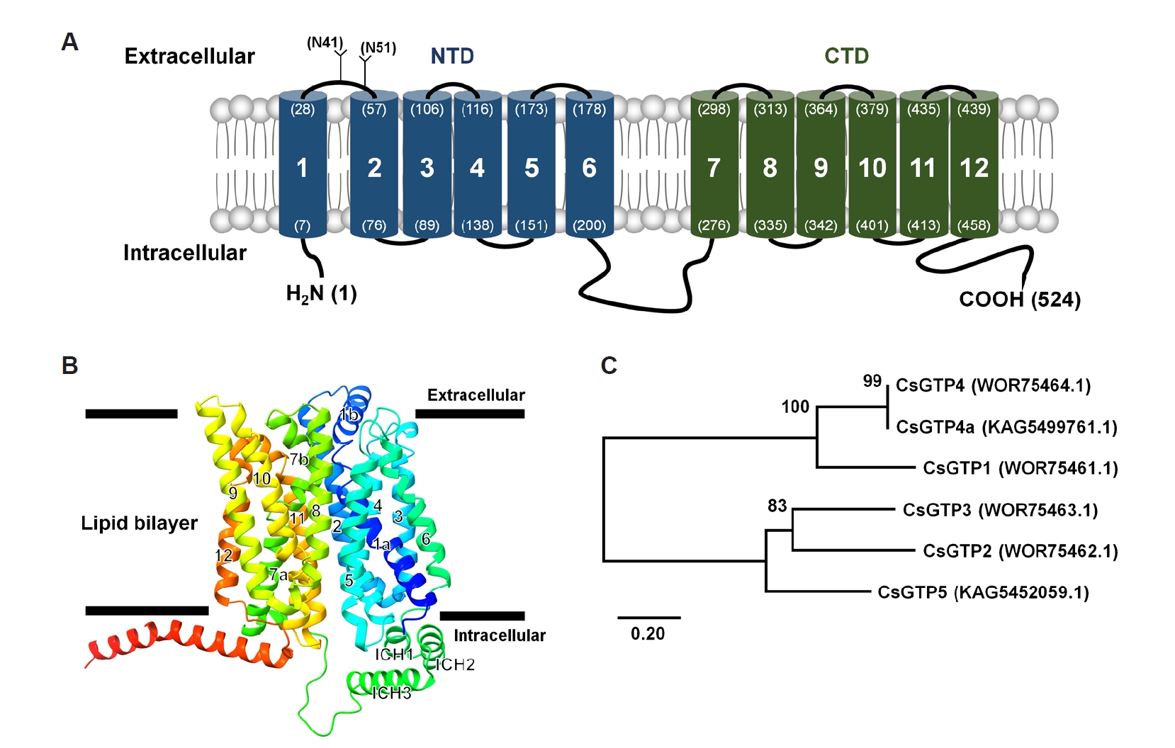
The open reading frame of CsGTP5 cDNA comprised 1,575 base pairs and encoded a protein of 524 amino acid residues, with a predicted molecular mass of 57.02 kDa. Topology analysis of the deduced amino acid sequence predicted 12 putative transmembrane domains, a characteristic feature of facilitative glucose transporter-related proteins (
Fig. 1A). Both the N- and C-termini were predicted to face the intracellular side of the plasma membrane (
Fig. 1A,
B).
Motif analysis identified 3 putative N-glycosylation motifs in CsGTP5. Based on the predicted membrane topology, 2 of these sites, located at residues 41 and 51, were positioned in the first extracellular loop between transmembrane domains 1 and 2, whereas the third site, located at residue 420, was predicted to lie within a transmembrane domain and is therefore unlikely to be glycosylated (
Supplementary Fig. S2). Phylogenetic analysis showed that CsGTP5 clustered with other glucose transporter-related proteins from
C. sinensis, supporting its classification as a member of the CsGTP family (
Fig. 1C).
To evaluate the substrate transport profile of CsGTP5, the uptake of several radiolabeled compounds was measured in
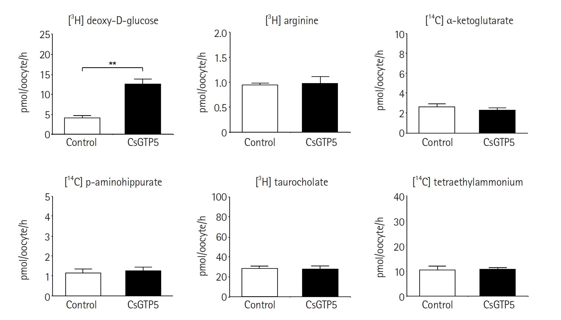
X. laevis oocytes expressing CsGTP5. Among the substrates examined, only [
3H] deoxy-D-glucose showed significantly increased uptake in CsGTP5-expressing oocytes. The uptake of [
3H] deoxy-D-glucose in CsGTP5-expressing oocytes was 12.7±3.9 pmol/oocyte/h, approximately 3.5-fold higher than that in water-injected control oocytes, which showed an uptake level of 3.6±1.5 pmol/oocyte/h (
Fig. 2). In contrast, no significant uptake of [
3H] arginine, [
14C] α-ketoglutarate, [
14C] p-aminohippurate, [
3H] taurocholate, or [
14C] tetraethylammonium was observed in CsGTP5-expressing oocytes (
Fig. 2). These results indicate that CsGTP5 preferentially mediates deoxy-D-glucose uptake among the tested substrates.
The transport properties of CsGTP5-mediated [
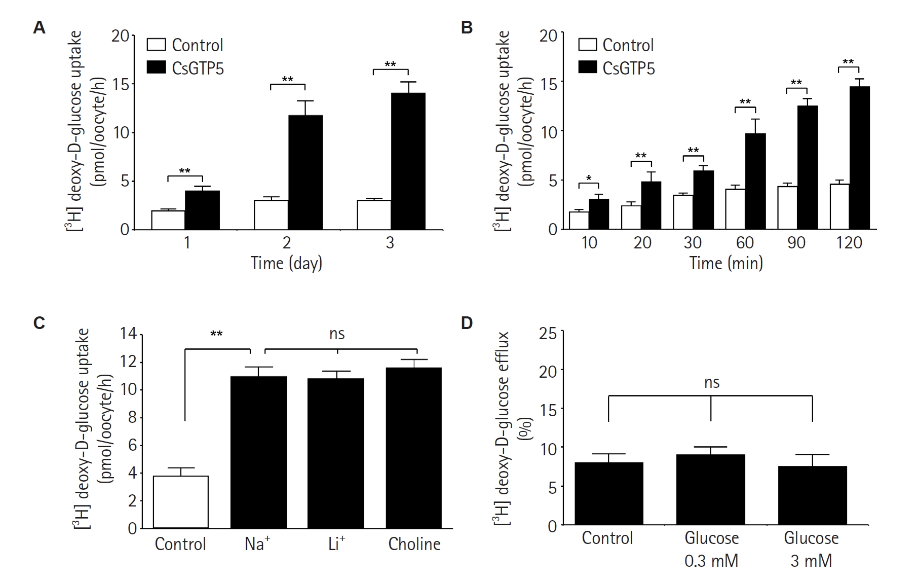
3H] deoxy-D-glucose uptake were further examined in oocytes. Uptake increased in an expression time-dependent manner from 1 to 3 days after cRNA injection (
Fig. 3A). In addition, [
3H] deoxy-D-glucose uptake increased with incubation time over the 10–120 min period (
Fig. 3B). These findings support the functional expression of CsGTP5 in the oocyte membrane and its role in mediating deoxy-D-glucose uptake.
To determine whether CsGTP5-mediated transport was dependent on extracellular sodium ions, Na
⁺ in the uptake buffer was replaced with an equivalent concentration of either Li
⁺ or choline. Replacement of Na
⁺ did not significantly affect [
3H] deoxy-D-glucose uptake during the 1 h incubation period (
Fig. 3C). These results suggest that CsGTP5-mediated deoxy-D-glucose uptake is not dependent on extracellular Na
⁺.
We next examined whether CsGTP5-mediated deoxy-D-glucose transport was trans-stimulated by extracellular glucose. The addition of unlabeled glucose at 0.3 or 3 mM, corresponding to 1,000-fold and 10,000-fold excess concentrations relative to [
3H] deoxy-D-glucose, did not significantly increase the efflux of preloaded [
3H] deoxy-D-glucose from CsGTP5-expressing oocytes (
Fig. 3D). This result suggests that extracellular glucose did not significantly stimulate CsGTP5-mediated [
3H] deoxy-D-glucose efflux under the experimental conditions tested.
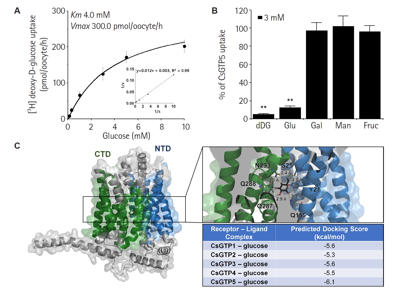
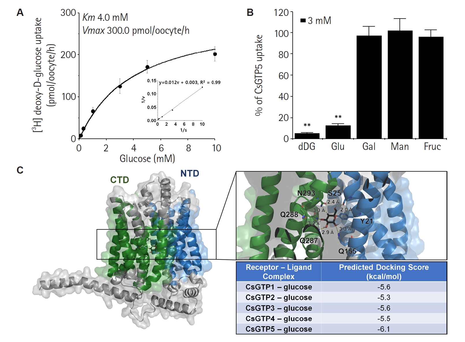
The concentration dependence of CsGTP5-mediated [
3H] deoxy-D-glucose uptake was analyzed to determine its kinetic properties. Uptake displayed saturable kinetics and was well fitted to the Michaelis–Menten equation (
Fig. 4A). Nonlinear regression analysis yielded an apparent
Km value of 4.0 mM and a
Vmax value of 300.0 pmol/oocyte/h. These kinetic properties indicate that CsGTP5 mediates carrier-dependent deoxy-D-glucose uptake.
To investigate the substrate selectivity of CsGTP5, competition assays were performed using unlabeled monosaccharides. The addition of 3 mM unlabeled deoxy-D-glucose or glucose strongly inhibited CsGTP5-mediated [
3H] deoxy-D-glucose uptake (
Fig. 4B). In contrast, galactose, mannose, and fructose did not show significant inhibitory effects. These results suggest that CsGTP5 preferentially recognizes deoxy-D-glucose and glucose over the other monosaccharides tested.
To further examine the structural plausibility of glucose recognition by CsGTP5, molecular docking analysis was performed using AutoDock Vina. Glucose was predicted to bind within the putative substrate-binding cavity of CsGTP5, where it formed predicted interactions with residues including Y21, S25, Q155, Q287, Q288, and N293 (
Fig. 4C). Comparative docking analysis of CsGTP1–5 showed predicted docking scores ranging from -5.3 to -6.1 kcal/mol. Among the 5 CsGTP proteins, CsGTP5 showed the lowest, but only slightly more favorable predicted docking score for glucose (
Fig. 4C). However, the differences among CsGTP docking scores were small, and these values should be interpreted as relative in silico predictions rather than experimental binding affinities.
Discussion
This study functionally characterized CsGTP5 as an additional glucose transporter-related protein derived from adult
C. sinensis. Previous studies have reported several glucose transport systems in
C. sinensis, including CsGLUT, CsGTP1–4, and CsSGLT [
3,
11,
12]. The identification of CsGTP5 expands this transporter repertoire and supports the idea that glucose transporter-related proteins in
C. sinensis may be functionally diverse rather than representing a single uniform glucose uptake pathway.
The predicted structure of CsGTP5 is consistent with that of facilitative glucose transporter-like proteins. CsGTP5 contains 12 putative transmembrane domains, a typical feature of major facilitator superfamily transporters, including members of the SLC2/GLUT family [
7,
8]. In addition, both the N-terminus and C-terminus were predicted to face the intracellular side of the membrane, further supporting its classification as a glucose transporter-related membrane protein [
23-
25]. The predicted extracellular N-glycosylation motifs may contribute to membrane localization, stability, or functional maturation, as reported for several mammalian GLUT proteins [
26,
27]. However, because glycosylation was not experimentally examined in this study, this remains a structural prediction.
Functionally, CsGTP5 shares several properties with previously characterized
C. sinensis glucose transporters. CsGTP5-mediated deoxy-D-glucose uptake was sodium-independent and saturable, which is consistent with facilitative glucose transport rather than sodium-coupled glucose transport [
7,
28]. Similar sodium-independent and saturable uptake has been reported for CsGLUT and CsGTP4 [
11,
12]. These similarities suggest that CsGTP5 belongs to the facilitative glucose transporter-like group of
C. sinensis transporters [
3].
At the same time, CsGTP5 appears to possess distinct transport characteristics. Its apparent
Km value suggests moderate affinity for deoxy-D-glucose, and its inhibition profile indicates selective recognition of deoxy-D-glucose and glucose rather than broad monosaccharide transport. This restricted substrate preference differs from CsGTP4, which has been reported to respond to glucose and galactose [
12]. These differences suggest that CsGTP proteins may not be functionally redundant, but instead may differ in substrate preference, affinity, and physiological role [
17,
29].
The absence of detectable glucose-stimulated efflux under the tested conditions further indicates that CsGTP5 did not show measurable exchange-like efflux activity in this assay. In some facilitative transporters, extracellular substrate can stimulate the efflux of preloaded intracellular substrate through exchange-like transport [
30,
31]. In the present study, extracellular glucose did not induce detectable efflux of preloaded [
3H] deoxy-D-glucose from CsGTP5-expressing oocytes under the tested conditions, suggesting that measurable exchange-like efflux activity was not observed in this assay. However, because the present assay measured cumulative efflux over 60 min and did not include higher extracellular glucose concentrations or shorter time-course measurements, these results do not fully exclude the possibility of rapid or concentration-dependent trans-stimulation. Further experiments will be required to clarify whether CsGTP5 exhibits exchange-like transport properties.
Molecular docking analysis provided structural support for the glucose-recognition capacity of CsGTP5. Glucose was predicted to bind within the putative substrate-binding cavity of CsGTP5, and CsGTP5 showed the lowest, although only slightly more favorable, predicted docking score among CsGTP1–5. However, the docking scores among CsGTP proteins were relatively close, and docking analysis cannot directly demonstrate binding affinity or transport activity [
32]. Therefore, the docking results should be interpreted as supportive and comparative in silico evidence that complements the functional uptake data.
Adult
C. sinensis survives for prolonged periods in the host bile duct and depends on host-derived nutrients for growth, survival, and reproduction [
2]. The presence of multiple glucose transporter-related proteins suggests potential functional diversity among transporter candidates involved in glucose acquisition. In this context, CsGTP5 may represent a functional glucose transporter-related candidate, as demonstrated by sodium-independent deoxy-D-glucose uptake in the heterologous oocyte expression system. However, because the present study was based on heterologous expression and did not include tissue localization, developmental expression, loss-of-function analysis, or parasite-based transport assays, the native physiological contribution of CsGTP5 to glucose uptake in adult worms remains to be determined. Although parasite nutrient transporters may be considered potential targets for future antiparasitic strategies, the physiological and therapeutic relevance of CsGTP5 will require further validation through tissue localization, developmental expression analysis, loss-of-function studies, and inhibitor-based transport assays [
9,
14].
In conclusion, CsGTP5 is a sodium-independent glucose transporter-related protein that mediates deoxy-D-glucose uptake. Its structural features, saturable transport kinetics, glucose-sensitive inhibition profile, and predicted glucose-binding capacity support its role as a facilitative glucose transporter-like protein. These findings provide additional evidence that glucose transporter-related proteins in C. sinensis may possess distinct transport properties within this transporter family.
Notes
-
Author contributions
Conceptualization: Cha SH, Han JH. Data curation: Cha SH, Lee WJ, Han JH. Formal analysis: Cha SH, Lee WJ, Syahada JH, Fitriana F, Rokhmad MF, Sim HY, Wu Z, Wang B, Han JH. Funding acquisition: Cha SH, Han JH. Investigation: Lee WJ, Syahada JH, Fitriana F, Rokhmad MF, Sim HY, Wu Z, Wang B. Methodology: Cha SH, Lee WJ, Han JH. Project administration: Cha SH, Han JH. Resources: Cha SH, Wang B, Han JH. Software: Cha SH, Wang B, Han JH. Supervision: Cha SH, Han JH. Validation: Cha SH, Wang B, Han JH. Visualization: Cha SH, Lee WJ, Syahada JH, Fitriana F, Rokhmad MF, Sim HY, Wu Z, Han JH. Writing – original draft: Cha SH, Lee WJ, Han JH. Writing – review & editing: Cha SH, Han JH.
-
Conflict of interest
Jin-Hee Han serves as an editor of Parasites, Hosts and Diseases but had no involvement in the decision to publish this article. No other potential conflicts of interest relevant to this study were reported.
-
Funding
This work was supported by the Inha Research Grant (SHC) and the National Research Foundation of Korea (NRF) funded by the Ministry of Science and ICT (MSIT) (RS-2025-16069701) (JHH).
-
Acknowledgments
Adult X. laevis was obtained from the Korean Xenopus Resource Center for Research.
Supplementary information
Fig. 1.Predicted structural features and phylogenetic relationship of CsGTP5. (A) Predicted membrane topology of full-length CsGTP5. CsGTP5 consists of 524 amino acids and contains 12 putative transmembrane domains. The predicted residue positions of the transmembrane domains are indicated in parentheses. NTD, N-terminal domain; CTD, C-terminal domain. (B) Predicted 3-dimensional structure of CsGTP5 generated using AlphaFold and visualized with UCSF ChimeraX. (C) Phylogenetic tree of CsGTP5 and selected glucose transporter-related proteins from Clonorchis sinensis. NCBI accession numbers are shown in parentheses.
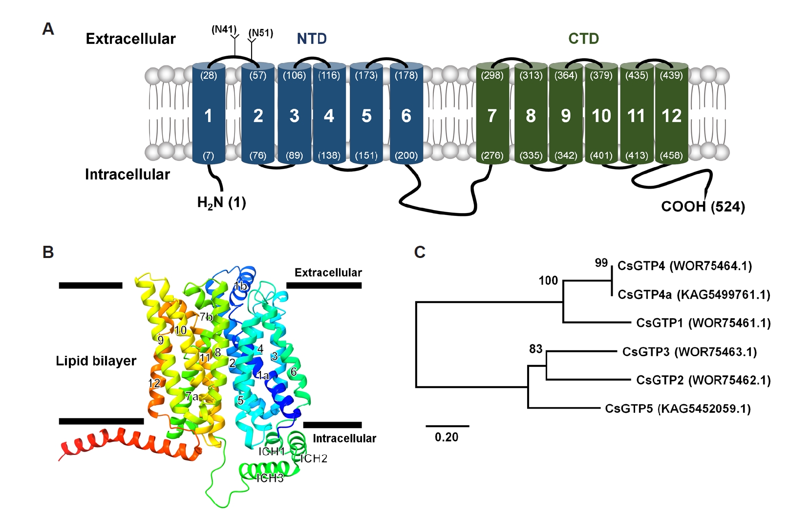
Fig. 2.Substrate uptake profile of CsGTP5 expressed in Xenopus laevis oocytes. Uptake of selected radiolabeled compounds was measured in water-injected control oocytes and CsGTP5-expressing oocytes for 1 h. The substrate concentrations were as follows: [3H] deoxy-D-glucose, 300 nM; [3H] arginine, 100 nM; [14C] α-ketoglutarate, 5 μM; [14C] p-aminohippurate, 10 μM; [3H] taurocholate, 200 nM; and [14C] tetraethylammonium, 10 μM. Data are presented as the mean±SE (n=6–8). **P<0.01 by Student t-test.
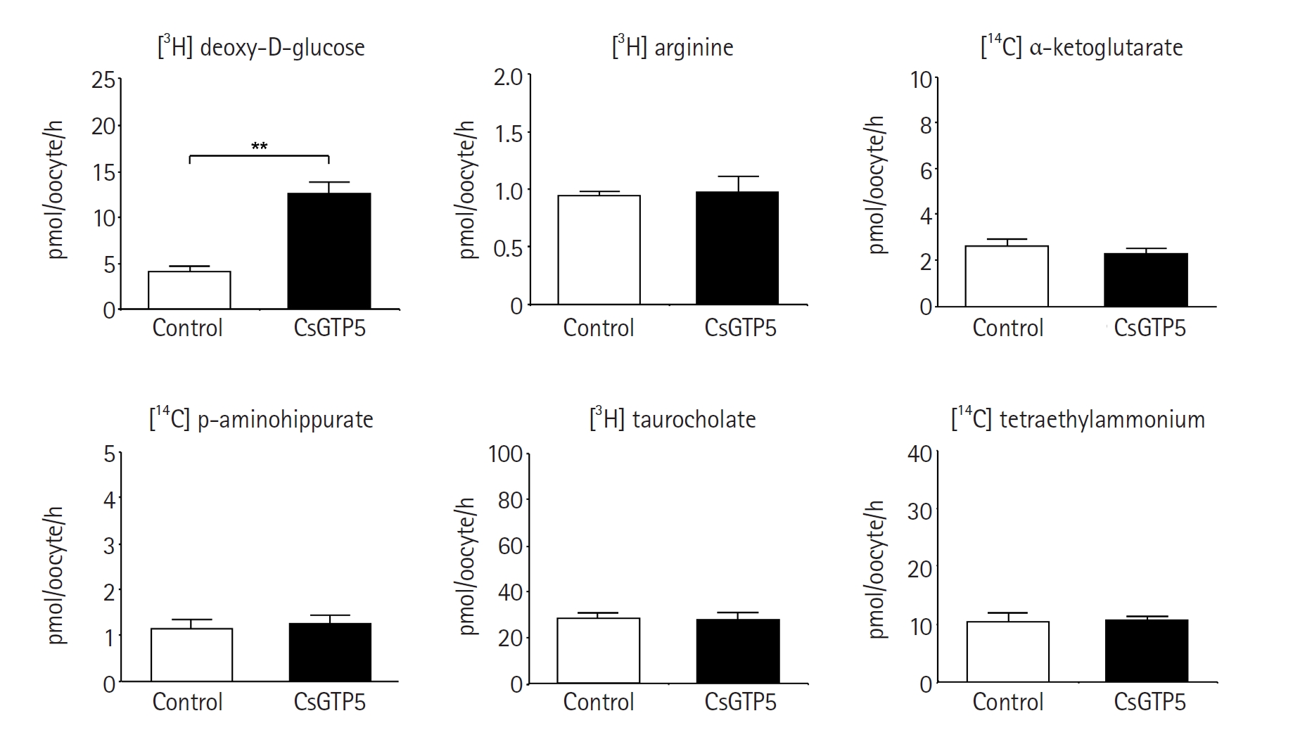
Fig. 3.Transport properties of CsGTP5-mediated [³H] deoxy-D-glucose uptake. (A) Expression time-dependent uptake of [3H] deoxy-D-glucose at 1, 2, and 3 days after CsGTP5 cRNA injection. (B) Incubation time-dependent uptake of [3H] deoxy-D-glucose over 10–120 min. (C) Effect of extracellular cation replacement on CsGTP5-mediated [3H] deoxy-D-glucose uptake. Extracellular Na⁺ was replaced with choline or Li⁺. (D) Efflux of preloaded [3H] deoxy-D-glucose from CsGTP5-expressing oocytes. Oocytes were preloaded with 300 nM [3H] deoxy-D-glucose for 90 min and incubated with or without unlabeled glucose at 0.3 or 3 mM for 60 min. Data are presented as the mean±SE (n=6–8). **P<0.01. ns, not significant.
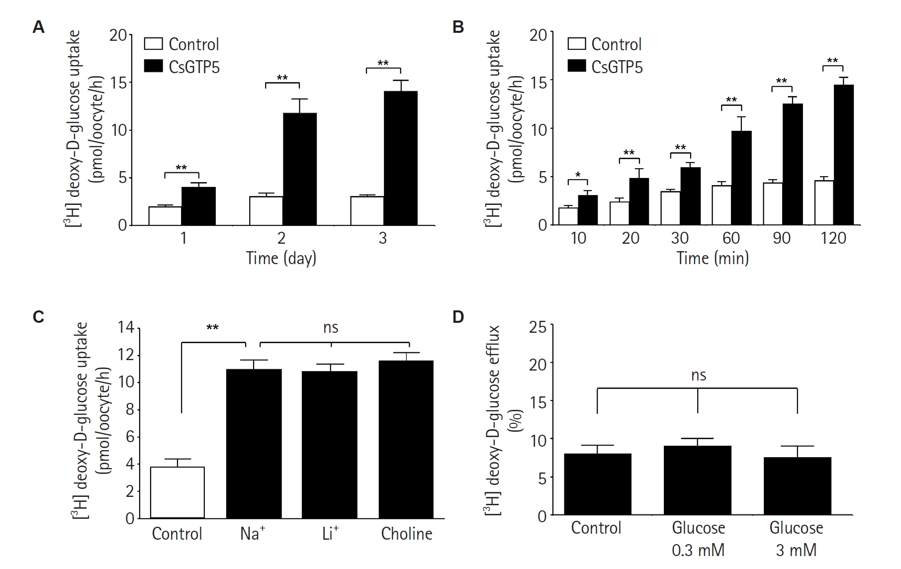
Fig. 4.Glucose transport properties and predicted glucose-binding features of CsGTP5. (A) Concentration-dependent uptake of [3H] deoxy-D-glucose mediated by CsGTP5. The inset shows Lineweaver–Burk analysis. V, velocity; S, concentration of deoxy-D-glucose. Data are presented as the mean±SE (n=6–8).(B) Effects of unlabeled monosaccharides on CsGTP5-mediated [3H] deoxy-D-glucose uptake. Oocytes were incubated with 300 nM [3H] deoxy-D-glucose in the absence or presence of 3 mM unlabeled monosaccharides. dDG, deoxy-D-glucose; Glu, glucose; Gal, galactose; Man, mannose; Fruc, fructose. Data are presented as the mean±SE (n=6–8). Statistical comparisons were performed against the no-competitor control. Connecting lines indicate the post hoc comparisons shown. **P<0.01. (C) Predicted glucose-binding pose in CsGTP5 and comparative AutoDock Vina docking scores for CsGTP1–5. CTD, C-terminal domain; NTD, N-terminal domain. Docking scores are shown as predicted binding energies in kcal/mol.
References
- 1. Qian MB, Keiser J, Utzinger J, Zhou XN. Clonorchiasis and opisthorchiasis: epidemiology, transmission, clinical features, morbidity, diagnosis, treatment, and control. Clin Microbiol Rev 2024;37:e0000923. https://doi.org/10.1128/cmr.00009-23
- 2. Tang ZL, Huang Y, Yu XB. Current status and perspectives of Clonorchis sinensis and clonorchiasis: epidemiology, pathogenesis, omics, prevention and control. Infect Dis Poverty 2016;5:71. https://doi.org/10.1186/s40249-016-0166-1
- 3. Dai F, Lee SO, Song JH, et al. Glucose transporters and sodium glucose co-transporters cooperatively import glucose into energy-demanding organs in carcinogenic liver fluke Clonorchis sinensis. PLoS Negl Trop Dis 2024;18:e0012315. https://doi.org/10.1371/journal.pntd.0012315
- 4. Wang X, Chen W, Huang Y, et al. The draft genome of the carcinogenic human liver fluke Clonorchis sinensis. Genome Biol 2011;12:R107. https://doi.org/10.1186/gb-2011-12-10-r107
- 5. You H, Stephenson RJ, Gobert GN, McManus DP. Revisiting glucose uptake and metabolism in schistosomes: new molecular insights for improved schistosomiasis therapies. Front Genet 2014;5:176. https://doi.org/10.3389/fgene.2014.00176
- 6. Skelly PJ, Tielens AG, Shoemaker CB. Glucose transport and metabolism in mammalian-stage schistosomes. Parasitol Today 1998;14:402-6. https://doi.org/10.1016/s0169-4758(98)01319-2
- 7. Mueckler M, Thorens B. The SLC2 (GLUT) family of membrane transporters. Mol Aspects Med 2013;34:121-38. https://doi.org/10.1016/j.mam.2012.07.001
- 8. Holman GD. Structure, function and regulation of mammalian glucose transporters of the SLC2 family. Pflugers Arch 2020;472:1155-75. https://doi.org/10.1007/s00424-020-02411-3
- 9. Krautz-Peterson G, Simoes M, Faghiri Z, et al. Suppressing glucose transporter gene expression in schistosomes impairs parasite feeding and decreases survival in the mammalian host. PLoS Pathog 2010;6:e1000932. https://doi.org/10.1371/journal.ppat.1000932
- 10. McKenzie M, Kirk RS, Walker AJ. Glucose uptake in the human pathogen Schistosoma mansoni is regulated through Akt/protein kinase B signaling. J Infect Dis 2018;218:152-64. https://doi.org/10.1093/infdis/jix654
- 11. Ahn SK, Cho PY, Na BK, et al. Molecular cloning and functional characterization of a glucose transporter (CsGLUT) in Clonorchis sinensis. Parasitol Res 2016;115:347-54. https://doi.org/10.1007/s00436-015-4754-y
- 12. Jun H, Mazigo E, Lee WJ, et al. Functional characterization of glucose transporter 4 involved in glucose uptake in Clonorchis sinensis. Parasites Hosts Dis 2024;62:450-60. https://doi.org/10.3347/PHD.24051
- 13. Lu Y, Yoo WG, Dai F, et al. Characterization of a novel organic solute transporter homologue from Clonorchis sinensis. PLoS Negl Trop Dis 2018;12:e0006459. https://doi.org/10.1371/journal.pntd.0006459
- 14. Dai F, Yoo WG, Lu Y, et al. Sodium-bile acid co-transporter is crucial for survival of a carcinogenic liver fluke Clonorchis sinensis in the bile. PLoS Negl Trop Dis 2020;14:e0008952. https://doi.org/10.1371/journal.pntd.0008952
- 15. Won JY, Louis JM, Roh ES, Cha SH, Han JH. Functional characterization of Clonorchis sinensis choline transporter. Parasites Hosts Dis 2023;61:428-38. https://doi.org/10.3347/PHD.23082
- 16. Lee WJ, Kim SJ, Lee WK, Han JH, Cha SH. Clonorchis sinensis dopamine transporter (CsDAT) facilitates dopamine uptake. Parasites Hosts Dis 2025;63:215-27. https://doi.org/10.3347/PHD.25040
- 17. Uldry M, Thorens B. The SLC2 family of facilitated hexose and polyol transporters. Pflugers Arch 2004;447:480-9. https://doi.org/10.1007/s00424-003-1085-0
- 18. Morrissey KM, Stocker SL, Wittwer MB, Xu L, Giacomini KM. Renal transporters in drug development. Annu Rev Pharmacol Toxicol 2013;53:503-29. https://doi.org/10.1146/annurev-pharmtox-011112-140317
- 19. Barghash A, Helms V. Transferring functional annotations of membrane transporters on the basis of sequence similarity and sequence motifs. BMC Bioinformatics 2013;14:343. https://doi.org/10.1186/1471-2105-14-343
- 20. Quistgaard EM, Low C, Guettou F, Nordlund P. Understanding transport by the major facilitator superfamily (MFS): structures pave the way. Nat Rev Mol Cell Biol 2016;17:123-32. https://doi.org/10.1038/nrm.2015.25
- 21. Drew D, North RA, Nagarathinam K, Tanabe M. Structures and general transport mechanisms by the major facilitator superfamily (MFS). Chem Rev 2021;121:5289-335. https://doi.org/10.1021/acs.chemrev.0c00983
- 22. Yamaoka K, Tanigawara Y, Nakagawa T, Uno T. A pharmacokinetic analysis program (multi) for microcomputer. J Pharmacobiodyn 1981;4:879-85. https://doi.org/10.1248/bpb1978.4.879
- 23. Mueckler M, Caruso C, Baldwin SA, et al. Sequence and structure of a human glucose transporter. Science 1985;229:941-5. https://doi.org/10.1126/science.3839598
- 24. Deng D, Xu C, Sun P. Crystal structure of the human glucose transporter GLUT1. Nature 2014;510(7503):121-5. https://doi.org/10.1038/nature13306
- 25. Deng D, Sun P, Yan C, et al. Molecular basis of ligand recognition and transport by glucose transporters. Nature 2015;526:391-6. https://doi.org/10.1038/nature14655
- 26. Breitling J, Aebi M. N-linked protein glycosylation in the endoplasmic reticulum. Cold Spring Harb Perspect Biol 2013;5:a013359. https://doi.org/10.1101/cshperspect.a013359
- 27. Asano T, Takata K, Katagiri H, et al. The role of N-glycosylation in the targeting and stability of GLUT1 glucose transporter. FEBS Lett 1993;324:258-61. https://doi.org/10.1016/0014-5793(93)80129-i
- 28. Gyimesi G, Pujol-Gimenez J, Kanai Y, Hediger MA. Sodium-coupled glucose transport, the SLC5 family, and therapeutically relevant inhibitors: from molecular discovery to clinical application. Pflugers Arch 2020;472:1177-206. https://doi.org/10.1007/s00424-020-02433-x
- 29. Burant CF, Bell GI. Mammalian facilitative glucose transporters: evidence for similar substrate recognition sites in functionally monomeric proteins. Biochemistry 1992;31:10414-20. https://doi.org/10.1021/bi00157a032
- 30. Carruthers A, DeZutter J, Ganguly A, Devaskar SU. Will the original glucose transporter isoform please stand up! Am J Physiol Endocrinol Metab 2009;297:E836-48. https://doi.org/10.1152/ajpendo.00496.2009
- 31. Vollers SS, Carruthers A. Sequence determinants of GLUT1-mediated accelerated-exchange transport: analysis by homology-scanning mutagenesis. J Biol Chem 2012;287:42533-44. https://doi.org/10.1074/jbc.M112.369587
- 32. Warren GL, Andrews CW, Capelli AM, et al. A critical assessment of docking programs and scoring functions. J Med Chem 2006;49:5912-31. https://doi.org/10.1021/jm050362n


 , Wang-Jong Lee2