Abstract
As gastroduodenoscopy performed more frequently, case reports of human echinostomiasis are increasing in Korea. A Korean woman presented at a local clinic with complaints of abdominal pain and discomfort that had persisted for 2 weeks. Under gastroduodenoscopy, two motile flukes were found attached on the duodenal bulb, and retrieved with endoscopic forceps. She had history of eating raw frog meat. The two flukes were identified as Echinostoma hortense by egg morphology, 27 collar spines with 4 end-group spines, and surface ultrastructural characters. This report may prove frogs to be a source of human echinostome infections.
-
Key words: Echinostoma hortense, duodenal bulb, gastroduodenoscopy, frog
INTRODUCTION
Echinostoma hortense is a member of the Echinostomatidae family, and is characterized by a large elongated body, a head crown with collar spines, a large oral sucker, tandem testes in the anterior body, and rich vitelline glands in the posterior body. The fluke inhabits the small intestine and prefers the upper region. Human echinostomatid infections, about 120 cases, have been reported in Korea. The echinostomatid species with a parasitologic history in Korean are
Echinostoma hortense,
E. cinetorchis,
Echinochasmus japonicus and
Acanthoparypium tyosenense (
Seo et al., 1983,
1985;
Ryang et al., 1985;
Chai et al., 1994,
2001). Of these
E. hortense is a dominant species in Korea. The mountainous villages of inland Korea represent its endemic foci (
Lee et al., 1988). Recently human
E. hortense infections were diagnosed by gastroduodenal endoscopy in Korea (
Chai et al., 1994;
Lee and Hong, 2002;
Cho et al., 2003). Here we describe a case of infection by
E. hortense in the duodenal bulb, which was diagnosed by upper endoscopy.
CASE RECORD
A Korean woman, 55-year-old, visited a local clinic with complaints of upper abdominal pains and discomfort on October 1999. Her symptoms had persisted for 2 weeks at the time of her visit. She lived in Ulsan-city, Gyeongsangnam-do, Korea. During gastroduodenal endoscopy, two motile flukes were found attached to the duodenal bulb. The flukes were slender, elongate, about 5 mm in length, with a red spot at anterior end (
Fig. 1). The flukes were removed from the duodenal bulb with endoscopic forceps. The patient was treated with single dose of praziquantel, 10 mg/kg. After treatment, the abdominal symptoms were subsided.
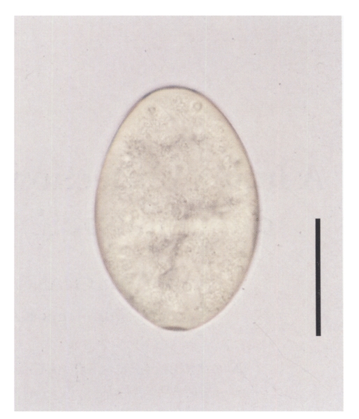
She had worked in a small restaurant, and had eaten raw frog meat several times about two months before onset of her symptoms. She denied ever eating freshwater fish in raw. Laboratory data were not available. A stool examination revealed six echinostome eggs, but none were found in family members. The eggs were bright yellow, elliptical, thin shelled with a shallow operculum, and measured 114.1 (111.2-118.6) µm long and 76.7 (74.1-79.0) µm wide (
Fig. 2).
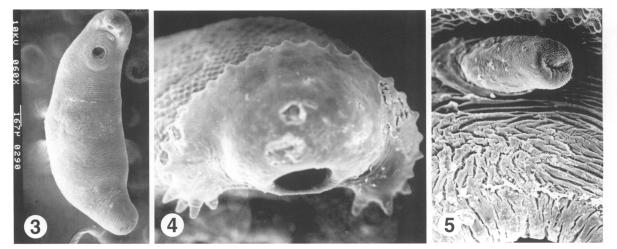
The flukes were processed for parasitological identification by light and scanning electron microscopic studies. The anterior of the fluke was bent ventrally at the acetabulum, and the surface was wrinkled transversely over the whole body. The body surface was covered with cobblestone-like cytoplasmic processes. Tegumental spines, scale-shaped with a broad base, were densely packed on the anterior surface of the body and then reduced in density and size on moving posteriorly (
Fig. 3). The anterior end bulged into a head crown around the oral sucker. The head crown was armed with uninterrupted 27 collar spines, of which the 4 end-group spines were present at the both ends of the the head crown (
Fig. 4). The outer surface of oral sucker was smooth and encircled with ciliated sensory (Type I) papillae arranged in 2-3 rows. Unciliated sensory (Type II) papillae, dome-like round structures, appeared on posterior half of the oral sucker lip. The acetabulum, wide-open, was aspinous with velvety cytoplasmic processes, and surrounded with Type I papillae. The cirrus erected from the genital opening revealed a velvety tegumental surface, and dense Type I sensory papillae, as compared with other parts of the body surface (
Fig. 5).
DISCUSSION
The number of collar and end-group spines is species-identifying morphological characteristic of echinostomatid flukes. The echinostomes retrieved from the present patient under scanning electron microscopy revealed 27 and 4 spines, which is a species-characteristic of
E. hortense. Surface ultrastructures, such as the arrangement of Type I sensory papillae on the oral sucker and around the acetabulum, and scale-shaped tegumental spines distributed at decreasing density posteriorly are consistent with previous descriptions of
E. hortense (
Lee et al., 1986b). After referring to surface ultrastructural characteristics and egg morphology (
Seo et al., 1983), the isolated flukes were identified as
E. hortense.
Adult
E. hortense inhabits the upper part of the small intestine in mammalian final hosts. The flukes produce ulcerative lesions in intestinal mucosa, villous atrophy characterized by blunting, villous fusion, and loss of villous tips, and crypt hyperplasia. In the stroma of villi were found inflammatory cell infiltration, vascular congestion, and edema (
Lee et al., 1990). The collar spines in the head crown of
E. hortense were stout and damaged the intestinal mucosa. Ulcerative lesions produced by adult
E. hortense in the duodenal bulb were previously observed by upper endoscopy (
Chai et al., 1994). In consideration of the pathologic findings, the symptoms experienced by the present patient, i.e., epigastric pain and abdominal discomfort, might be provoked by the abrasive action of
E. hortense. The frequent symptoms reported by infected individuals, though not pathogen-specific, are abdominal pain, cramps, anorexia, postprandial burning, flatulence, and diarrhea (
Lee et al., 1986a;
Chai et al., 1994;
Lee and Hong, 2002). It has also been suggested that chronic and severe infection may cause intestinal villous atrophy and lead to manifestations of malabsorption.
Freshwater fish have been recorded as infection sources of echinostomiasis hortense, most frequently loaches in Korea, followed by frogs (
Chai et al., 1985;
Ryang et al., 1985;
Cho et al., 2003). Frog tadpoles become infected with metacercariae of echinostomes and metamorphose to adult frogs with the metacercariae in their tissues, predominantly in muscles. Metacercariae were previously found mainly in the dorsal pharyngeal wall, body cavity, and mesentery of
Rana nigromaculata (
Rim et al., 1982). People may become infected by eating inadequately cooked frog meat, as occurred in the present case. The reservoir hosts have been recognized to play a significant role in an ecosystem of digenean trematodes. They initiate the trematode's life cycle by supplying trematode eggs to the first intermediate molluscan hosts. Of reservoir hosts, rats and dogs are reported to show a high prevalence of
E. hortense infection in Korea (
Seo et al., 1981). This human case reports that frogs are a source of human echinostome infections in Korea.
References
- 1. Chai JY, Han ET, Park YK, Guk SM, Lee SH. Acanthoparyphium tyosenense: the discovery of human infection and identification of its source. J Parasitol 2001;87:794-800.
- 2. Chai JY, Hong SJ, Sohn WM, Lee SH, Seo BS. Studies on intestinal trematodes in Korea XVI. Infection status of loaches with the metacercariae of Echinostoma hortense. Korean J Parasitol 1985;23:18-23.
- 3. Chai JY, Hong ST, Lee SH, Lee GC, Min YI. A case of echinostomiasis with ulcerative lesions in the duodenum. Korean J Parasitol 1994;32:201-204.
- 4. Cho CM, Tak WY, Kweon YO, Kim SK, Choi YH, Kong HH, Chung DI. A human case of Echinostoma hortense (Trematoda: Echinostomatidae) infection diagnosed by gastroduodenal endoscopy in Korea. Korean J Parasitol 2003;41:117-120.
- 5. Lee OJ, Hong SJ. Gastric echinostomiasis diagnosed by endoscopy. Gastrointest Endosc 2002;55:440-442.
- 6. Lee SK, Chung NS, Ko IH, Ko HI, Chai JY. Two cases of natural human infection by Echinostoma hortense. Korean J Parasitol 1986a;24:77-81. (in Korean).
- 7. Lee SK, Chung NS, Ko IH, Sohn WM, Hong ST, Chai JY, Lee SH. An epidemiological survey of Echinostoma hortense infection in Chongsong-gun, Kyungbuk Province. Korean J Parasitol 1988;26:199-206. (in Korean).
- 8. Lee SH, Hong SJ, Chai JY, Hong ST, Seo BS. Tegumental ultrastructures of Echinostoma hortense observed by scanning electron microscopy. Korean J Parasitol 1986b;24:63-70. (in Korean).
- 9. Lee SH, Noh TY, Sohn WM, Kho WG, Hong ST, Chai JY. Chronological observation of intestinal lesions of rats experimentally infected with Echinostoma hortense. Korean J Parasitol 1990;28:45-52. (in Korean).
- 10. Rim HJ. Echinostomiasis. CRC Handbook Series in Zoonoses, Section C: Parasitic Zoonoses. 1982. Vol. III (Trematode Zoonoses):Boca Raton, Florida, USA. CRC Press, Inc.; p. 53-69.
- 11. Ryang YS, Ahn YK, Lee KW, Kim TS, Han MH. Two cases of natural human infection by Echinostoma hortense and its second intermediate host in Wonju area. Korean J Parasitol 1985;23:33-40. (in Korean).
- 12. Seo BS, Cho SY, Hong ST, Hong SJ, Lee SH. Studies on parasitic helminthes of Korea. V. Survey on intestinal trematodes of house rats. Korean J Parasitol 1981;19:131-136.
- 13. Seo BS, Chun KS, Chai JY, Hong SJ, Lee SH. Studies on intestinal trematodes in Korea XVII. Development of egg lying capacity of Echinostoma hortense in albino rats and human experimental infection. Korean J Parasitol 1985;23:24-32.
- 14. Seo BS, Hong ST, Chai JY, Lee SH. Studies on intestinal trematodes in Korea VIII. A human case of Echinostoma hortense infection. Korean J Parasitol 1983;21:219-223.
Fig. 1Echinostomes (arrows) on the duodenal bulb.
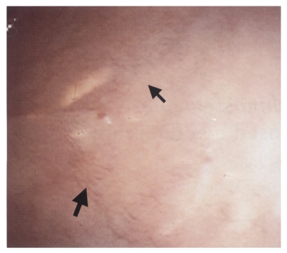
Fig. 2An egg of Echinostoma hortense from the patient. Bar = 54 µm.

Fig. 3-5Scanning electron micrographs of an Echinostoma hortense specimen retrieved from the patient. Fig. 3. Ventral view. Fig. 4. The oral opening and head crown spikes with collar spines. Fig. 5. The cirrus erected from the genital pore anterior to the acetabulum.

Citations
Citations to this article as recorded by

- Clonorchis sinensis and Echinostoma hortense detected by endoscopy and molecular characterization: two case reports and update on diagnosis
Lijia Wen, Benhe Wang, Hui Zhang
Frontiers in Medicine.2025;[Epub] CrossRef - Rare Case of Echinostoma cinetorchis Infection, South Korea
Sooji Hong, Hyejoo Shin, Yoon-Hee Lee, Sung-Jong Hong, So-Ri Kim, Youn-Kyoung Kim, Young-Jin Son, Jeong-Gil Song, Jong-Yil Chai, Bong-Kwang Jung
Emerging Infectious Diseases.2024;[Epub] CrossRef - Type of cercaria in freshwater snails at Tunggu Pampang Reservoir, Makassar City, Indonesia
Arif Rahman Jabal, Dian Mutiasari, Hairil Akbar, M. Arfah, Marhani Marhani, Rini Rini, Nur Alam Sobak, Anggit Julianingsih Pisu, Agnes Immanuela Toemon, Arini Ratnasari
Russian Journal of Infection and Immunity.2023; 12(4): 765. CrossRef - Neglected food-borne trematodiases: echinostomiasis and gastrodiscoidiasis
Rafael Toledo, María Álvarez-Izquierdo, J. Guillermo Esteban, Carla Muñoz-Antoli
Parasitology.2022; 149(10): 1319. CrossRef - Morphology and Molecular Identification of Echinostoma revolutum and Echinostoma macrorchis in Freshwater Snails and Experimental Hamsters in Upper Northern Thailand
Preeyaporn Butboonchoo, Chalobol Wongsawad, Pheravut Wongsawad, Jong-Yil Chai
The Korean Journal of Parasitology.2020; 58(5): 499. CrossRef - Fasciola hepatica infection in children actively detected in a survey in rural areas of Mardan district, Khyber Pakhtunkhawa province, northern Pakistan
Asma W. Qureshi, Aurang Zeb, Abu Mansoor, Azam Hayat, Santiago Mas-Coma
Parasitology International.2019; 69: 39. CrossRef - Infection Status of Isthmiophora hortensis Metacercariae in Dark Sleepers, Odontobutis Species, from Some Water Systems of the Republic of Korea
Woon-Mok Sohn, Byoung-Kuk Na, Shin-Hyeong Cho, Jung-Won Ju
The Korean Journal of Parasitology.2018; 56(6): 633. CrossRef - New Definitive Hosts and Differential Body Indices of Isthmiophora hortensis (Digenea: Echinostomatidae)
Woon-Mok Sohn, Byoung-Kuk Na, Sung-Shik Shin
The Korean Journal of Parasitology.2017; 55(3): 287. CrossRef - Echinostoma macrorchis (Digenea: Echinostomatidae): Metacercariae in Cipangopaludina chinensis malleata Snails and Adults from Experimental Rats in Korea
Woon-Mok Sohn, Byoung-Kuk Na
The Korean Journal of Parasitology.2017; 55(5): 541. CrossRef - Epidemiological analysis of human fascioliasis in northeastern Punjab, Pakistan
Asma W. Qureshi, Akhtar Tanveer, Santiago Mas-Coma
Acta Tropica.2016; 156: 157. CrossRef - An update on human echinostomiasis
R. Toledo, J. G. Esteban
Transactions of The Royal Society of Tropical Medicine and Hygiene.2016; 110(1): 37. CrossRef - A Case of Echinostoma cinetorchis (Trematoda: Echinostomatidae) Infection Diagnosed by Colonoscopy
Woon Tae Jung, Kyeong Ju Lee, Hong Jun Kim, Tae Hyo Kim, Byoung-Kuk Na, Woon-Mok Sohn
The Korean Journal of Parasitology.2014; 52(3): 287. CrossRef - Echinostoma hortense Infection with Enteritis Diagnosed by Upper Gastrointestinal Endoscopy in a Dog
Hiroki OKANISHI, Jun MATSUMOTO, Sadao NOGAMI, Yumiko KAGAWA, Toshihiro WATARI
Journal of Veterinary Medical Science.2013; 75(7): 991. CrossRef - Evaluation of the role of rats as reservoir hosts for fishborne zoonotic trematodes in two endemic northern Vietnam fish farms
Nguyen Lan Anh Thi, Henry Madsen, Dao Thanh Ha, Eric Hoberg, Anders Dalsgaard, K. Darwin Murrell
Parasitology Research.2012; 111(3): 1045. CrossRef - Proteomics of foodborne trematodes
Rafael Toledo, M. Dolores Bernal, Antonio Marcilla
Journal of Proteomics.2011; 74(9): 1485. CrossRef - Echinostoma hortense and Heterophyid Metacercariae Encysted in Yellowfin Goby, Acanthogobius flavimanus, from Shinan-gun and Muan-gun (Jeollanam-do), Korea
Woon-Mok Sohn, Byoung-Kuk Na, Shin-Hyeong Cho
The Korean Journal of Parasitology.2009; 47(3): 307. CrossRef - Foodborne Intestinal Flukes in Southeast Asia
Jong-Yil Chai, Eun-Hee Shin, Soon-Hyung Lee, Han-Jong Rim
The Korean Journal of Parasitology.2009; 47(Suppl): S69. CrossRef - ECHINOSTOMA HORTENSE ASADA INFECTION IN THE DUODENUM: INCIDENTAL FINDINGS DURING ROUTINE GASTROINTESTINAL ENDOSCOPY
Toshiaki Tanaka, Tomoari Kamada, Hideki Koga, Aki Tanaka, Noriaki Manabe, Jiro Hata, Hiroaki Kusunoki, Manabu Ishii, Motonori Sato, Kenichi Tarumi, Akiko Shiotani, Tetsuya Okino, Ken Haruma
Digestive Endoscopy.2008; 20(2): 87. CrossRef - Eosinophilia: Causes and pathobiology in persons with prior exposures in tropical areas with an emphasis on parasitic infections
Yae-Jean Kim, Thomas B. Nutman
Current Infectious Disease Reports.2006; 8(1): 43. CrossRef