Abstract
Trichinellosis is a parasitic zoonosis of public health importance. It is caused by Trichinella spiralis which has a wide host range including humans. In the present communication, the ELISA technique was employed on a total of 803 blood samples from 7 selected pig breeding farms in 1996 for diagnosis and surveillance of trichinellosis. Out of the entire 803 samples, nine were found to be suspected while one was positive by ELISA. But western blot analyses employed for further confirmation have shown that all of 10 samples did not react to larval excretory-secretory product antigens. These results indicate that pig breeding farms included in the present study are free from trichinellosis. However, it does not mean Korea is free from trichinellosis since human trichinellosis has recently been reported. The necessity of continued surveillance for trichinellosis in both pigs and wild animals was discussed.
-
Key words: Trichinella spiralis, enzyme-linked immunosorbent assay, western blotting, swine, Korea
Trichinellosis is a parasitic zoonosis infecting both humans and animals, although infection rate in domestic animals is invariably low. There have been no reported cases of trichinellosis in domestic pigs in Korea, but there were cases of
Trichinella spiralis infection in people who had consumed raw liver, spleen, blood and muscle of a badger (
Sohn et al., 2000). This incidence may indicate that Korea is no longer free from trichinellosis.
Pigs are considered to be the major source of Trichinella infection for humans. As a matter of fact, we heavily rely on the imported pigs for breeding; therefore, it is possible that the parasite has already been introduced to Korea. In the present report, the authors have applied the enzyme-linked immunosorbent assay (ELISA) technique for the serological surveillance and the quarantine of the selected pig breeding farms in Korea. ELISA confirmed positive samples were then subjected to the western blot analysis for further confirmation.
Trichinella spiralis had been routinely maintained in the laboratory by serial passages through the artificial infection in rats (Sprague-Dawley) and pigs. Larval excretory-secretory product (ESP) was obtained for the antigen. For the preparation of the positive antiserum, three pigs were orally inoculated with T. spiralis larvae (L3, 20,000/piglet). Two pigs not inoculated were used as positive controls. Negative sera were also obtained from the three pigs immediately before inoculation. After inoculation they were kept separately in individual stalls.
The blood samples from the three pigs infected with T. spiralis were subjected to ELISA, as described previously by Office International des Epizooties (Manual of standards for diagnostic tests & vaccines, 2000. OIE). Briefly, T. spiralis excretory-secretory antigens were diluted to 5 µg/ml in coating buffer (50 mM carbonate/bicarbonate, pH 9.6). The 96-well microtitre plates were coated with the antigens (100 µl/well) at 4℃ overnight. The wells were washed 3 times with phosphate buffered saline containing 0.5% Tween 20 (PBS-Tween). And then the following reagents (100 µg/ml) were added to the wells sequentially and incubated for 30 minutes at room temperature between each step; swine serum (diluted 1:100 in PBS-Tween); rabbit anti-swine IgG (diluted 1:1,000 in PBS-Tween); goat anti-rabbit IgG-peroxidase conjugate (diluted 1:1,000 in PBS-Tween). Bound enzyme was visualized by the addition of 5'-aminosalicylic acid (0.8 µg/ml) with 0.005% hydrogen peroxide (pH 5.6-6.0).
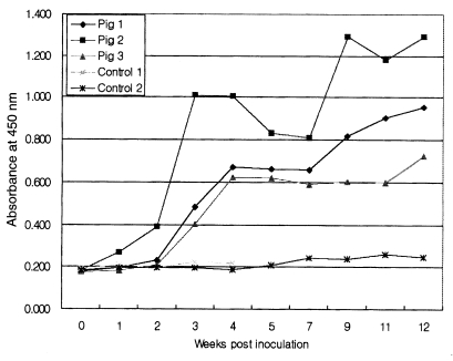
The optical densities (OD) increased remarkably at week 3 post-inoculation (PI) and the positive/negative ratios were estimated as 3.5 to 7.5 throughout the experimental period as shown in
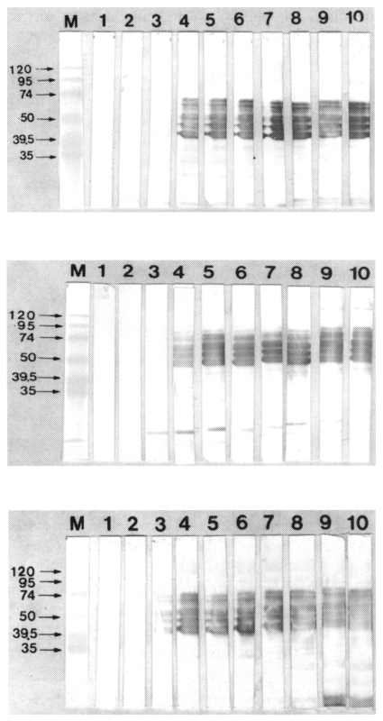
Fig. 1. The same blood samples were then tested by western blot (WB) at weekly and/or biweekly intervals. WB analyses have shown that antibodies reacted to the sera at week 3 PI, specifically the 50 kDa protein group (
Fig. 2). The ESP antigens recognized in worm secretions consisted of a group of structurally related glycoproteins with molecular weights between 45 and 55 kDa (
Gamble and Graham, 1984). Serological tests were performed on 803 sera from seven selected pig breeding farms for swine trichinellosis (
Table 1).
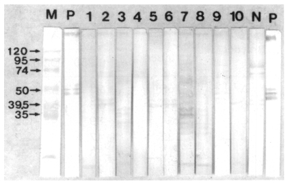
Of 803 sera tested, 9 samples (1.1%) were ELISA suspected (OD > 0.625) while only one sample (0.1%) was ELISA positive (OD > 0.75). According to international standard (OIE 2000), the values 4 times that of the normal serum pool controls are considered to be positive and the values 3 times higher than the normal are classified as the suspected. To eliminate false-positive reactions in the ELISA, 10 samples (ELISA suspected and positive) were analyzed by WB. The results have shown that all of the samples did not react to the ESP antigens (
Fig. 3). These results indicate that pig breeding farms included in the present study are free from trichinellosis. Most of serum samples showed low level ELISA values in these farms. It may indicate that no more infection or reinfection activities of
T. spiralis. The results do not mean, however, that Korea is free from
Trichinella infection. There have been human trichinellosis cases reported recently in Korea (
Sohn et al., 2000). All of the cases involved the consumption of raw badger meat. Although the source of the infection is not yet known, it indicates that the life cycle of
T. spiralis is maintained in the wildlife in Korea.
The authors suggest that it is necessary to continue an intensive national level surveillance for trichinellosis in both domestic pigs and wild animals in Korea. The OIE international animal health code (
2000) recommends a trichinellosis surveillance program in which the serological surveys should be conducted statistically every 5 years on slaughtered pig population sufficient to provide at least 95% confidence level.
The advantage of serological diagnosis is the increased sensitivity over the direct demonstration methods, and the serological responses in pigs persisted for at least 6 months PI with no decline (
Gamble et al., 1983;
Gamble et al., 1988;
Gamble, 1996). In slaughtering inspection, the ELISA yielded less than 0.3% false-positives and was nearly 100% sensitive in detecting infected pigs with more than 1 larva per gram of tissue (
Seawright et al., 1983;
Oliver et al., 1989). The serological testing is of particular value for both the surveillance of trichinellosis and the slaughtering inspection. Although the ELISA has yielded promising results, false-positive and false-negative reactions with swine sera have not been completely eliminated due to the crude nature of the antigen preparation, especially in false-positive reactions. Thus, the researchers have attempted to purify
Trichinella antigens with the use of monoclonal antibodies (
Gamble and Graham, 1984). The results in the present communication indicate that the combination of ELISA screening and WB confirmation system is a good technique for large-scale epidemiological studies and serological surveillance of trichinellosis.
ACKNOWLEDGEMENT
We would like to thank Professor S. Y. Cho and H. W. Nam, Department of Parasitology, Catholic University of Korea, for kindly providing rats infected with Thai strain of Trichinella spiralis.
References
- 1. Gamble HR. Detection of trichinellosis in pigs by artificial digestion and enzyme immunoassay. J Food Prot 1996;59:295-298.
- 2. Gamble HR, Anderson WR, Graham CE, Murrell KD. Diagnosis of swine trichinosis by enzyme-linked immunosorbent assay (ELISA) using an excretory-secretory antigen. Vet Parasitol 1983;13:349-361.
- 3. Gamble HR, Graham CE. Monoclonal antibody-purified antigen for the immunodiagnosis of trichinosis. Am J Vet Res 1984;45:67-74.
- 4. Gamble HR, Rapic D, Marinculic A, Murrell KD. Evaluation of excretory-secretory antigens for the serodiagnosis of swine trichinellosis. Vet Parasitol 1988;30:131-137.
- 5. Office International des Epizooties. Trichinellosis. OIE Manual of standards, List B Disease. 2000. Chapter 3.5.3.
- 6. Oliver DG, Singh P, Allison DE, et al. In: Tanner CE, editor. Field evaluation of an enzyme immunoassay for detection of hogs in a high volume north Carolina abattoir. Trichinosis. 1989. Madrid, Spain. Consejo Superior de investigaciones Press; p. 439-444.
- 7. Seawright GL, Despommier D, Zimmerman W, Isenstein RS. Enzyme immunoassay for swine trichinellosis using antigens purified by immunoaffinity chromatography. Am J Trop Med Hyg 1983;32:1275-1284.
- 8. Sohn WM, Kim HM, Chung DI, Yee ST. The first human case of Trichinella spiralis infection in Korea. Korean J Parasitol 2000;38:111-115.
Fig. 1Serum antibody (IgG) response of pigs experimentally infected with Trichinella spiralis larvae (L3, 20,000/piglet 1, 2 and 3) by ELISA.

Fig. 2Enzyme Immuno-transfer blotting of excretory-secretory antigens detected with specific sera from three experimentally infected pigs. M, marker proteins; Lane 1, the sera from 3 pigs before the inoculation; Lanes 2-10, the sera from 3 pigs after the inoculation 1, 2, 3, 4, 5, 7, 9, 11, 12 weeks, respectively.

Fig. 3Enzyme Immuno-transfer blotting of excretory-secretory antigens detected with 1 sero-positive and 9 suspected samples (optical density > 0.625) in field trials. M, marker proteins; P, positive sample; N, nagative sample; Lanes 1-9, suspected samples; Lane 10, sero-positive sample.

Table 1.Distribution of ELISA values against Trichinella spiralis in pigs from 7 breeding farms in Korea
Table 1.
|
Breeder |
Heads examined |
Absorbance value (405 nm)
|
|
≤ 0.250 |
0.251-0.375 |
0.376-0.50 |
0.501-0.625 |
0.626-0.750 |
≥ 0.751 |
|
A |
58 |
23 |
16 |
12 |
5 |
2 |
- |
|
B |
63 |
26 |
23 |
12 |
- |
1 |
1 |
|
C |
254 |
89 |
132 |
25 |
5 |
3 |
- |
|
D |
132 |
47 |
42 |
22 |
18 |
3 |
- |
|
E |
110 |
53 |
50 |
7 |
- |
- |
- |
|
F |
58 |
27 |
31 |
- |
- |
- |
- |
|
G |
128 |
50 |
69 |
9 |
- |
- |
- |
|
|
Total |
803 |
315 |
363 |
87 |
28 |
9 |
1 |
Citations
Citations to this article as recorded by

- Prevalence of Trichinella spp. antibodies in wild boars (Sus scrofa) and domestic pigs in Korea
H.J. Kim, W.S. Jeong, E.M. Kim, S.G. Yeo, D.J. An, H. Yoon, E.J. Kim, C.K. Park
Veterinární medicína.2015; 60(4): 181. CrossRef - Primary characterization and assessment of a T. spiralis antigen for the detection of Trichinella infection in pigs
Aleksandar Zocevic, Sandrine A. Lacour, Pauline Mace, Baldissera Giovani, Aurelie Grasset-Chevillot, Isabelle Vallee, Pascal Boireau
Veterinary Parasitology.2014; 205(3-4): 558. CrossRef - Evaluation of ELISA coupled with Western blot as a surveillance tool for Trichinella infection in wild boar (Sus scrofa)
Leigh Cuttell, Maria Angeles Gómez-Morales, Beth Cookson, Peter J. Adams, Simon A. Reid, Paul B. Vanderlinde, Louise A. Jackson, C. Gray, Rebecca J. Traub
Veterinary Parasitology.2014; 199(3-4): 179. CrossRef - Seroprevalence of trichinellosis in domestic animals in northwestern Vietnam
N. Vu Thi, N.V. De, N. Praet, L. Claes, S. Gabriël, P. Dorny
Veterinary Parasitology.2013; 193(1-3): 200. CrossRef - Development and evaluation of an immunochromatographic strip for trichinellosis detection
Gai-Ping Zhang, Junq-Qing Guo, Xuan-Nian Wang, Jun-Xing Yang, Yan-Yan Yang, Qing-Mei Li, Xue-Wu Li, Rui-Guang Deng, Zhi-Jun Xiao, Ji-Fei Yang, Guang-Xu Xing, Dong Zhao
Veterinary Parasitology.2006; 137(3-4): 286. CrossRef