Abstract
The present study analyzed serum IgG subclass antibody reaction to major antigenic bands of Clonorchis sinensis to investigate improvement of its serodiagnosis. Of the four subclass antibodies, IgG1 and IgG2 antibodies were produced but not specific, IgG3 antibody was least produced, and IgG4 antibody was prominent and specific. The serum IgG antibody reaction to any of 43-50, 34-37, 26-28, and 8 kDa bands was found in 65.5% of 168 egg positive cases while IgG4 antibody reaction was found in 22.0% of them. The positive rates of IgG and IgG4 antibodies were directly correlated with the intensity of infection. All of the sera from heavily infected cases over EPG 5,000 showed positive reaction for specific IgG and IgG4 antibodies. The specific serum IgG4 antibody disappeared within 6 months after treatment. The bands of 35 kDa and 67 kDa cross-reacted with IgG antibodies but not with IgG4 antibodies in sera of other trematode infections. The present findings suggest that serum IgG4 antibody reaction to 8 kDa band is specific but not sensitive. Any method to increase its sensitivity is required for improved serodiagnosis.
-
Key words: clonorchiasis, serodiagnosis, IgG, IgG4, 8 kDa antigen
INTRODUCTION
Clonorchis sinensis Looss, 1907 is one of trematodes of the human bile duct which is widely prevalent in East Asia including Korea, China, Russia, and Vietnam, and about 28 millions of the cases are estimated in China (
Li, 1997). Clonorchiasis is the most prevalent helminthiasis in Korea as the egg positive rate was 1.4% in 1997 throughout the country (
Ministry of Health and Welfare and Korea Association of Health, 1997). Its prevalence in Korea was rather high in 1971 as 4.6% but gradually and continuously decreased thereafter. The gradual decrease has been mainly induced by the government-supported control program with praziquantel treatment and health education but also to water pollution. The decrease of clonorchiasis in Korea is very slow compared to that of other parasite infections. Still one million cases of clonorchiasis are estimated in Korea and adequate control strategy is essential to minimize its medical and social impacts.
Screening of subjected population at the field and detection of the infected cases is the beginning point of its control. Fecal examination is the standard diagnostic method until now, but collection of feces becomes more and more difficult at the field because of indifference of the inhabitants. Furthermore collection and examination of feces requires much labor and time, which makes the field work of large scale difficult. Serological screening by ELISA or other techniques is a candidate to replace the fecal examination because serological screening can be executed together with other serological or hematological examinations (
Rim, 1990;
Yong et al., 1991).
Since
C. sinensis is a lumen-dwelling parasite, serological reaction by ELISA is not so strong enough resulting in low sensitivity except in cases of heavy infection (
Hong, 1988). A study revealed low specificity of serological diagnosis in clonorchiasis because of cross-reaction and residual reaction after cure (
Hong et al., 1997). The serological studies have used crude antigen and observed reactions of total IgG antibodies in serum. To make better diagnostic efficacy of serology, it is essential to analyze the antigens and the antibody reactions in detail. The present study applied immunoblotting and observed the serum IgG subclass antibody reactions to several antigenic bands by infection intensity and after cure.
MATERIALS AND METHODS
Preparation of C. sinensis antigen
Metacercariae of C. sinensis were collected from naturally infected Pseudorasbora parva by pepsin digestion and orally infected to New Zealand white rabbits. Adult worms of C. sinensis were recovered from the liver of the rabbits 3 months later, and homogenized in phosphate-buffered saline (PBS, pH 7.4). After high speed centrifugation (15,000 rpm for 1 hr), the supernatant was used as soluble crude extract antigen, and aliquots of 0.2 ml (1 mg/ml) were stored at -70℃ until use.
Sera
One hundred sixty eight sera were collected from C. sinensis egg positive cases by fecal examination and 75 sera from C. sinensis egg negative cases. The fecal examination was done by both modified Kato-Katz method and formalin-ether sedimentation technique. All of the egg positive cases were treated with praziquantel and some of their sera were collected 6 months after treatment.
For screening of cross-reaction, 14 sera of Paragonimus westermani, 21 of Metagonimus yokogawai, 12 of Gymnophalloides seoi, 1 of Fasciola hepatica, 9 of sparganum, and 24 of Taenia solium cysticercus infections were obtained from the positive cases confirmed by fecal examination or multi-antigen ELISA.
SDS-PAGE and immunoblotting
Protein bands of crude
C. sinensis antigen were separated under reducing conditions by SDS-PAGE on 7.5-15% polyacrylamide gels and transferred to PVDF membrane as previously described (
Hong et al., 1997). The membrane was cut into strips and each strip was incubated overnight with 1:100 diluted human serum at room temperature. Peroxidase-conjugated anti-human IgG goat serum (whole molecule specific; Cappel, Cochranville, PA, USA) was used after 1:1,000 dilution for IgG antibody reaction. For IgG subclass antibody reactions, HRP-conjugated anti-human IgG1, IgG2, IgG3, and IgG4 mouse sera (Southern Biotechnology Associates, Inc., Birmingham, USA) were used as 1:1,000 diluted. The blots were developed with 0.03% 4-chloro-1-naphthol containing 0.03% H
2O
2, in PBS (0.01 M, pH 7.4).
Statistical significance was evaluated by the LOGISTIC procedure. A P-value less than 0.05 was considered significant.
RESULTS
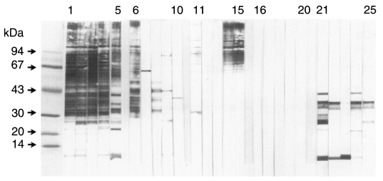
Immunoblotting showed strong reactions of IgG and IgG4 antibodies in cases of clonorchiasis to 43-50, 34-37, 26-28, and 8 kDa bands while no reaction was recognized with serum of normal controls. Many bands of large molecules reacted with IgG1 and IgG2 antibodies but not specific, and no IgG3 antibody reacted to the bands (
Fig. 1). Of the four bands, 34-37 kDa band showed major antigenic reaction with serum IgG antibody; in 52.6% of 137 cases with EPG under 500, in 78.9% of 19 cases with EPG 600-5,000, and in 100% of 12 cases with EPG over 5,100 (
Table 1). However, the 35 and 67 kDa bands showed cross-reaction with serum IgG antibodies of other trematode infections while IgG4 subclass antibodies showed no reaction (
Table 2;
Fig. 2).
As for the reaction with IgG4 antibody, 8 kDa band was the major antigen; 5.8% of the cases under EPG 500, 57.9% of the cases with EPG 600-5,000, and 91.7% of the cases over EPG 5,100 (
Table 1). There was no cross reaction of the 8 kDa band with serum IgG4 antibody of other trematode infections (
Fig. 2).
Immunoblotting demonstrated that the reactions to 26-28 kDa and 8 kDa bands disappeared 6 months after treatment (
Fig. 3).
DISCUSSION
The present study showed IgG4 subclass antibodies were predominantly produced in clonorchiasis, and IgG1 and IgG2 subclass antibodies were also produced but IgG3 were not (
Fig. 1). Prominence of IgG4 antibodies is already well-known in several kinds of human helminthiases, such as schistosomiasis (
Hakangard et al., 1996), hookworm disease (
Loukas et al., 1996), hydatid disease (
Aceti et al., 1993), and cysticercosis (
Yang et al., 1998). In clonorchiasis, increase of specific serum IgG, IgE and IgA antibodies is already known (
Yen et al., 1992;
Yong et al., 1993;
Lin et al., 1995;
Hong et al., 1997), but IgG antibody is the major antibody in serum.
The IgG4 subclass antibody produced in clonorchiasis was demonstrated more specific than IgG1 and IgG2 subclass antibodies. The IgG1 and IgG2 subclass antibodies were produced more in serum than IgG4 antibodies, but most of them reacted to several non-specific bands. Furthermore, the IgG2 subclass antibody was still maintained even 6 months after cure. Therefore these IgG1 and IgG2 subclass antibodies are related with background serological reaction, which makes the serology of clonorchiasis with crude antigen less specific.
Hong et al. (
1997) observed that 43, 34, and 25-28 kDa bands are specific antigenic bands in human clonorchiasis, and Kim (
1998) recorded that 7 kDa band is specific for active clonorchiasis. The present findings re-confirm that major antigenic molecules of
C. sinensis are 43-50, 34-37, 26-28, and 8 kDa bands. These bands reacted to both IgG antibodies and IgG4 subclass antibodies (
Fig. 1). The 7 kDa band of Kim (
1998) and the present 8 kDa band are looking same, but of different size estimation. The nature of the band must be characterized by further studies.
The case frequency of positive reaction differs by the antigenic bands (
Table 1). For IgG antibody reaction, 34-37 kDa band showed 58.9% frequency of 168 egg positive cases and is the most frequent one. Whole positive rate to any of the four bands was 65.5%. For IgG4 antibody reaction, the frequency to individual antigenic bands was much lower than that of IgG antibody reaction which means screening of IgG4 antibodies is very low of its sensitivity (
Table 1).
The level of specific serum IgG antibody is known to correlate with EPG counts (
Hong, 1988), and the present findings also show very close correlation between the immunoblotting reaction frequency and EPG counts. The whole frequency of IgG antibody reaction was 60.6% in cases of EPG fewer than 500, but it increased to 100% in cases of EPG over 5,100 (
Table 1). The frequency of IgG4 antibody reaction also increased by EPG, as it was 8.8% in cases of EPG fewer than 500 but 100% in those of EPG over 5,100. This finding suggests that biased selection of the cases makes quite different results of serology. Therefore, EPG counts of subjected cases should be considered for evaluation of clonorchiasis serology. Since most of clonorchiasis cases in Korea are light infected (
Ministry of Health and Welfare and Korea Association of Health, 1997), low sensitivity of its serology should be overcome for proper serodiagnosis. Otherwise, serodiagnosis is of no value.
It was reported that serum IgG antibodies significantly decreased within 3 to 7 months after treatment with praziquantel and IgA antibodies decreased within 1 month (
Hong, 1988;
Lin et al., 1995). The present study showed that IgG and IgG4 antibodies to 26-28 and 8 kDa bands disappeared within 6 months after treatment. Specific serum antibodies evidently decreased within a few months after cure. Especially the serological reaction to 26-28 and 8 kDa bands may be a marker of active infection.
Cross-reaction was found between the 35 or 67 kDa antigenic bands and serum IgG antibodies of other trematode or cestode infections, such as
P. westermani,
M. yokogawai,
G. seoi and cysticercus of
T. solium (
Table 2,
Fig. 2). The 35 and 67 kDa bands are certainly shared with other trematodes or cestodes, and are the major molecule for cross-reaction in serological diagnosis of clonorchiasis. For better specificity of serological diagnosis, it is essential to exclude these two bands in preparation of the antigen. The antigen cocktail of 26-28 and 8 kDa antigenic bands with or without 43-50 and 34-37 bands may be very useful for serodiagnosis of clonorchiasis.
In conclusion, the present finding confirms that detection of IgG4 antibody is less sensitive but more specific than detection of IgG antibody in serodiagnosis of clonorchiasis.
Notes
-
This study was supported by a grant from 1995-1997 National R&D program (MOST), Korea.
ACKNOWLEDGEMENTS
We thank Professor Seung-Yull Cho, Department of Molecular Parasitology, Sungkyunkwan University, School of Medicine for kind supply of human sera of other helminth infections.
References
- 1. Aceti A, Pennica A, Teggi A, et al. IgG subclasses in human hydatid disease: prominence of the IgG4 Response. Int Arch Allergy Immunol 1993;102:347-351.
- 2. Hakangard C, Deelder AM, Gabone RM, Nilsson LA, Ouchterlony O. A comparative study on specific antibodies and circulating antigen (CAA) in serum and parasitological findings for diagnosis of schistosomiasis mansoni in an endemic area in Tanzania. Acta Trop 1996;61:213-222.
- 3. Hong ST. Changes of anti-Clonorchis sinensis IgG antibody in serum after praziquantel treatment in human clonorchiasis. Korean J Parasitol 1988;26:1-8.
- 4. Hong ST, Kho WG, Lee M, Lee JS, Lee SH. Immunoblot patterns of clonorchiasis. Korean J Parasitol 1997;35:87-93.
- 5. Kim SI. A Clonorchis sinensis-specific antigen that detects active human clonorchiasis. Korean J Parasitol 1998;36:37-45.
- 6. Li B, Wang E, Chao Y, Chao Y. Clonorchis sinensis and clonorchiasis. 1997. Shenyang. Shenyang Publication Co.; (in Chinese).
- 7. Lin YL, Chen ER, Yen CM. Antibodies in serum of patients with clonorchiasis before and after treatment. Southeast Asian J Trop Med Public Health 1995;26:114-119.
- 8. Loukas A, Opdebeeck J, Croese J, Prociv P. Immunoglobulin G subclass antibodies against excretory/secretory antigens of Ancylostoma caninum in human enteric infections. Am J Trop Med Hyg 1996;54:672-676.
- 9. Ministry of Health and Welfare and Korea Association of Health. Prevalence of intestinal parasitic infections in Korea. - The Sixth Report -. 1997. Seoul.
- 10. Rim HJ. Clonorchiasis in Korea. Korean J Parasitol 1990;28(suppl):63-78.
- 11. Yang HJ, Chung JY, Yun DH, et al. Immunoblot analysis of a 10 kDa antigen in cyst fluid of Taenia solium metacestodes. Parasite Immunol 1998;20:483-488.
- 12. Yen CM, Chen ER, Hou MF, Chang JH. Antibodies of different immunoglobulin isotypes in serum and bile of patients with clonorchiasis. Ann Trop Med Parasitol 1992;86:263-269.
- 13. Yong TS, Im K, Chung PR. Analysis of Clonorchis sinensis antigens and diagnosis of clonorchiasis using monoclonal antibodies. Korean J Parasitol 1991;29:293-310.
- 14. Yong TS, Kim DS, Lee SY, Im K, Lee KY. Detection of specific serum IgE in clonorchiasis cases and analysis of Clonorchis sinensis allergens. Yonsei Med J 1993;34:248-257.
Fig. 1Immunoblot analysis of IgG antibodies and IgG subclass antibodies of Clonorchis sinensis infected sera. Lanes 1-5, IgG antibodies; lanes 6-10, IgG1; lanes 11-15, IgG2; lanes 16-20, IgG3; lanes 21-25, IgG4 subclass antibodies.

Fig. 2Cross-reaction with sera of other parasite infections. A. IgG antibody reaction. B. IgG4 antibody reaction. lanes 1-3, cysticercus of Taenia solium; lanes 4-17, Paragonimus westermani; lanes 18-24, Gymnophalloides seoi; lane 25, sparganum; lane 26, Fasciola hepatica. The 35 and 67 kDa bands are reacting to serum IgG antibodies of other parasite infections but no bands reacted to IgG4 antibodies.
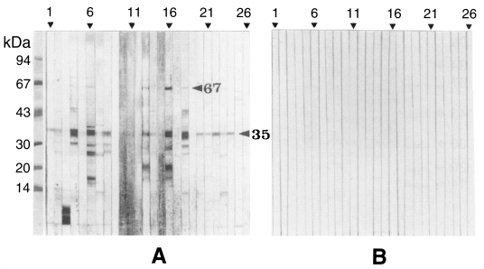
Fig. 3Immunoblot analysis of serum IgG and IgG4 antibodies of Clonorchis sinensis infected cases, paired samples after treatment. Lanes 1-10, IgG antibodies in paired sera pre- (odd numbers) and post-treatment (even numbers) of 5 cases; lanes 11-20, IgG4 antibodies in paired sera pre- (odd numbers) and post-treatment (even numbers) of 5 cases.
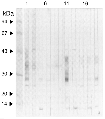
Table 1.Number and proportion (%) of serum IgG and IgG4 antibody positive cases to antigenic
fractions of Clonorchis sinensis by EPG grade
Table 1.
|
EPG grade |
No. of cases |
|
No. (%) of positive casesa) to bands |
|
43-50 kDa |
34-37 kDa |
26-28 kDa |
8 kDa |
Total |
|
0-500 |
137 |
IgG |
16 (11.7) |
72 (52.6) |
21 (15.3) |
20 (14.6) |
83 (60.6) |
|
|
IgG4 |
4 (2.9) |
8 (5.8) |
6 (4.4) |
8 (5.8) |
12 (8.8) |
|
600-5000 |
19 |
IgG |
7 (36.8) |
15 (78.9) |
11 (57.9) |
14 (73.7) |
15 (78.9) |
|
|
IgG4 |
5 (26.3) |
10 (52.6) |
8 (42.1) |
11 (57.9) |
13 (68.4) |
|
5100- |
12 |
IgG |
12 (100) |
12 (100) |
12 (100) |
12 (100) |
12 (100) |
|
|
IgG4 |
10 (83.3) |
10 (83.3) |
10 (83.3) |
11 (91.7) |
12 (100) |
|
Total |
168 |
IgG |
35 (20.8) |
99 (58.9) |
44 (26.2) |
46 (27.4) |
110 (65.5) |
|
|
IgG4 |
19 (11.3) |
28 (16.7) |
24 (14.3) |
30 (17.9) |
37 (22.0) |
Table 2.Reaction number and rate (%) of serum IgG antibodies of other parasite infections to 35 kDa and 67 kDa antigen bands of Clonorchis sinensis
Table 2.
|
Sera of infection with |
No. of examined |
No. (%) of positive cases to |
|
35 kDa |
67 kDa |
|
Paragonimus westermani
|
14 |
7 (50.0) |
5 (35.7) |
|
Metagonimus yokogawai
|
21 |
15 (71.4) |
4 (19.0) |
|
Gymnophalloides seoi
|
12 |
6 (50.0) |
1 (8.3) |
|
Fasciola hepatica
|
1 |
0 (0) |
0 (0) |
|
sparganum |
9 |
0 (0) |
0 (0) |
|
cysticercus of Taenia solium
|
24 |
11 (45.8) |
4 (16.7) |