Abstract
After morphological grouping of Acanthamoeba by Pussard and Pons, phylogeny of the genus has been always a big topic to the researchers. Because of the variability of morphological characteristics, unchangeable and stable characters have been investigated for phylogenic criteria. Isoenzyme and mitochondrial DNA RFLP (Mt DNA RFLP) analyses revealed different patterns among strains assigned to a same species. Therefore, these characteristics would be considered as tools for strain discrimination than species identification. The most recently developed and the most promising method is the sequence analysis of 18s ribosomal RNA coding DNA (18s rDNA). The phylogenic tree based on comparison of 18s rDNA sequences distinguished the 3 morphological groups of Acanthamoeba and divided them into 12 unique sequence types (T1-T12 genotypes). Most clinical and environmental isolates belonged to the morphological group II and the genotype T4. In the Republic of Korea, 2 strains of Acanthamoeba, YM-2 and YM-3, were first isolated from the environment in 1974. However, phylogenic identification of Korean Acanthamoeba isolates from human infections or the environment were tried from the late 1990s. By RFLP analysis or total sequence analysis of 18s rDNA revealed that almost all clinical isolates including the one from a suspicious granulomatous amebic encephalitis patient belonged to the genotype T4. A large number of environmental isolates from contact lens storage cases, tapped water, and ocean sediments also belonged to the genotype T4. Almost identical strain characteristics, such as Mt DNA RFLP pattern of environmental isolates, with the clinical isolates could make a simple conclusion that most environmental isolates might be a potential keratopathogen.
-
Key words: Acanthamoeba, molecular phylogeny, mitochondrial DNA, 18s rDNA
INTRODUCTION
The genus
Acanthamoeba Volkonsky, 1931 has adapted to live in a variety of human environments, such as soil, air, freshwater, swimming pools, tap water, and ocean sediments. Recently, medical protozoologists have paid attention to
Acanthamoeba since several species of the genus have been known to cause life-threatening granulomatous amebic encephalitis (GAE) in immunocompromised individuals and sight-threatening amebic keratitis in contact lens wearers. The incidence of
Acanthamoeba keratitis infection has increased exponentially during the last 30 years [
1,
2]. Up to the year 2009, 2 suspicious GAE cases were found to be infected with
Acanthamoeba spp. in the Republic of Korea [
3,
4]. These patients died of meningoencephalitis. In addition, 1 case of
Acanthamoeba pneumonia with an immunodeficiency status was reported [
4]. In the case of
Acanthamoeba keratitis, the incidence is increasing from the early 1990s and more than 30 cases were reported already.
Despite the medical importance of the genus, identification and classification of acanthamoebae at the subgenus level has been problematic. The identification of
Acanthamoeba at the generic level can be easily accomplished by the morphological characteristics. Pussard and Pons [
5] divided the genus into 3 morphological groups according to the cyst size and other morphological features. Group 1 consists of
Acanthamoeba spp. with relatively large cysts, distinctly stellate endocysts, and smooth spherical ectocysts. Group 2 and group 3
Acanthamoeba spp. have smaller cysts (less than 18 µm in diameter). Species in Group 2 have polygonal to stellate endocysts with irregular or wrinkled ectocysts, while the cysts of group 3 species have rounded or slightly angular endocysts with tinner and smooth or slightly wrinkled ectocysts. The grouping has been widely used before species identification of the amoeba. However, the morphologic characteristics of the cyst can change with the culture conditions [
6,
7] and highly variable within the same strain (
Fig. 1). Therefore, species identification by morphology alone can hardly be possible [
8].
Investigators have used several kinds of non-morphological methods for the taxonomy of Acanthamoeba spp. Among the methods, isoenzyme analysis, RFLP of mitochondrial DNA (Mt DNA), RFLP of small subunit ribosomal DNA (ssu rDNA), and the sequence analysis of ssu rDNA have been recently applied and regarded promising. In this paper, the potential of these molecular methods for the phylogeny of Acanthamoeba, and the molecular phylogeny of Acanthamoeba korean isolates, were briefly reviewed.
ISOENZYME ANALYSIS AND MITOCHONDRIAL DNA RFLP
Isoelectric focusing of isoenzymes had been applied for identification of
Acanthamoeba spp. Studies of isoenzyme patterns [
9-
12] suggested the other groupings of
Acanthamoeba strains that are not consistent with previous species assignment based on the morphological criteria of Pussard and Pons [
5]. De Jonckheerez [
13] examined 30 strains of 15
Acanthamoeba species for the isoenzyme patterns by agarose isoelectric focusing (IEF) in the pH range 3-10. Comparing zymograms of 5 enzymes, he reassigned the strains of amoebae to each species. Kong et al. [
14] observed the interstrain polymorphism of the isoenzyme profiles among 7 strains of
Acanthamoeba morphologically assigned to
Aanthamoeba polyphaga. Since considerable variation of zymograms was noted within a species of
Acanthamoeba, isoenzyme study alone is not regarded fully sufficient for species identification.
Mt DNA RFLP analysis has also been applied for taxonomy of
Acanthamoeba. Bogler et al. [
15] prospected the availability of Mt DNA RFLP for the identification of
Acanthamoeba after analysis of 13 strains. They observed that most of the
Acanthamoeba strains had distinct fragment patterns of Mt DNA. Yagita and Endo [
16] also analyzed 8 isolates which had been identified as
Acanthamoeba castellaniii or A. polyphaga, and reported variability of Mt DNA digestion phenotypes among different strains of
A. castellanii. Kong et al. [
14] confirmed the variability in digestion phenotype of
A. polyphaga as well as that of
A. castellanii. As for the interstrain sequence diversity of Mt DNA of
Acanthamoeba, other investigators reported similar results [
15,
17-
19].
Considering profound interstrain diversity in a species of the Mt DNA RFLP and alloenzyme profiles, Kong et al. [
14] and Chung et al. [
20] suggested that both analyses should be used for strain identification, differentiation, and characterization rather than species identification.
SMALL SUBUNIT RIBOSOMAL RNA CODING DNA RFLP (RIBOPRINTING)
PCR-based DNA restriction analyses of 18s rDNA (riboprinting) of
Acanthamoeba showed promising results for species differentiation of
Acanthamoeba [
21]. The sequence of primers for PCR was designed to hybridize at the highly conserved sequences at the 5'and 3'termini of eukaryotic ssu rDNA [
22]. They analyzed 23 reference strains of
Acanthamoeba, including 18 (neo) type strains for classification at the subgenus level by riboprinting. The dendrogram based on the riboprinting coincided well with the grouping of Pussard and Pons [
5] based on the morphological features of the cysts. Khan and Paget [
23] applied similar methods to identify clinical and environmental isolates from UK. They amplified a part of 18S rDNA with genus-specific primers [
23] and digested the products with restriction enzymes. The riboprinting can be applied for rapid identification of
Acanthamoeba isolated from the clinical and environmental specimens under limited condition to obtain the DNA sequence of the samples.
18S rDNA SEQUENCE ANALYSIS
The most recently proposed method for molecular taxonomy of
Acanthamoeba is 18s rDNA sequence analysis. Byers group investigated the 18s rRNA gene phylogeny [
24,
25]. After the examination of the 18s rDNA sequence variation in a group of 53 strains that includes the morphologically assigned 18 isolates and 35 new isolates, they suggested 12 sequence types (T1-T12). The sequence types were defined as the sequences or groups of sequences that differ from all other sequences by at least 6%. In the phylogeny by 18s rDNA sequence analysis, 3 morphological groups were clearly distinguished. Each of the morphological groups consisted of several different lineages of sequence types. Three species in the morphological group I were identified as T7 for
Acanthamoeba astronyxis, T8 for
Acanthamoeba tubiashi, and T9 for
Acanthamoeba comandoni. In group 3,
Acanthamoeba culbertsoni was identified as T10 and
A. healyi as T12, and
Acanthamoeba palestinensis,
Acanthamoeba pustulosa, and
Acanthamoeba lenticulata represented T1, T2, and T5, respectively. The other 3 types, T3, T4, and T11 are included in group 2; T3 for
Acanthamoeba griffinii and T11 for
Acanthamoeba hatchetti and
Acanthamoeba stevensoni. The biggest lineage T4 contains all the remaining group 2 isolates. However, by this system, the relationship among all taxa within T4 was not clearly defined. Strains in T4 are too closely related to be resolved fully by use of 18s rDNA sequences. They proposed that each sequence type should be equated with a single species. The various species in T4 might be all reclassified as
A. castellanii, because the sequence type includes the type strain for that species.
Although phylogeny by 18s rDNA sequence analysis has a limitation to distinguish species in sequence type T4, it is practical to classify clinical or environmental samples for species identification. Now the identification of a new isolate of
Acanthamoeba from clinical sample or environment is performed mainly by 18s rDNA sequence analysis [
26-
29].
With the 18s rDNA sequence analysis, reassignment of several Acanthamoeba spp. originally assigned by morphological criteria have been suggested. Chung's group evaluated the taxonomic validity of 4 species of Acanthamoeba; A. divionensis, A. paradivionensis, A. mauritaniensis, and A. rhysodes and suggested that 3 species should be regarded as synonyms of A. rhysodes. They exhibited 18s rDNA sequence differences of 0.2-1.1 with each other.
Since most clinical or environmental isolates belong to 18s rDNA sequence type T4, many investigators prefer to classify an isolate as their genotypes than scientific names even in the case of already named species, such as A. castellanii Castellani. It is necessary and urgent to define the strains in the genotype T4 as a species complex or as several species based on other molecular characteristics.
MITOCHONDRIAL 16S rDNA SEQUENCE ANALYSIS
The value of mitochondrial small subunit rRNA gene (mt ssu rDNA) analysis as a taxonomic tool for
Acanthamoeba isolates with close interrelationship was also evaluated [
30,
31]. Mt DNA is a molecular clone within mitochondriate eukaryotic cells like
Acanthamoeba. Merit of Mt ssu rDNA is lack of intron, and smaller and more consistent in size than nuclear ssu rDNA. Yu et al. [
30] compared the phylogeny based on the PCR-RFLP pattern of mt ssu rDNA with that of genomic ssu rDNA PCR-RFLP. Ledee et al. [
31] analyzed Mt ssu rDNA sequences of 68 isolates and constructed a phylogenetic tree. The similar genotype clustering by Mt ssu rDNA analysis with that by nuclear ssu rDNA genes indicated that those clustering represent the main branches in the phylogeny of
Acanthamoeba. Moreover, based on the subgrouping of T4 genotypes into 8 clades, they suggested that Mt ssu rDNA sequence may serve as the best available molecular basis for differentiating the species with this genotype.
Acanthamoeba PHYLOGENY IN KOREA
It was middle of the 1970s that the first suspicious
Acanthamoeba meningoencephalitis case was reported in the Republic of Korea [
3]. The patient was a 5-year-old boy who developed multiple subcutaneous, non-tuberculous granulomas in many organs, and died of meningoencephalitis. However, unfortunately the organism identification was not performed. It was also in the middle of the 1970s that the first strain of
Acanthamoeba was isolated in Korea [
32].
Acanthamoeba spp., YM-2, YM-3, and YM-4, were isolated from water puddles, a storing reservoir, and the gill of fresh water fish. However, the phylogenic positioning of those isolates was tried quite long time later because of lack of standardized species identification system.
The second case of
Acanthamoeba meningoencephalitis in Korea was a 7-year-old boy with facial laceration near eyebrow by accident.
Acanthamoeba trophozoites were observed in his skin biopsy after his death [
4]. However, species identification of the causative
Acanthamoeba was tried after 13 years later based on 18s rDNA partial sequence analysis. For partial amplification, 2 sets of genus-specific primers were designed to hybridize to the specific regions of 18S rDNA of the genus
Acanthamoeba (
Fig. 2). The primer sets, VR1 and VR2, encompassing the variable regions of
A. castellanii Castellani 18S rDNA were designed to hybridize from 111-326 and 822-1,090 nucleotide numbers, respectively. The sequences of these primers were as follows: VR1F (TTATAGTTTATTTGATGGTC), VR1R (TGCGATCCGAAAGA-GTTACT), VR2F (CCGTCCCCTCCTTCTGGATT) and VR2R (CTATCCCTATT-AATCATTAC). Two fragments, sized 200 and 270 bp, of the variable region of 18s rDNA was amplified by PCR from the paraffin block of the skin biopsy. Combined sequences of 2 variable regions were subjected to construct phylogenetic tree by comparison with that of reference strains (
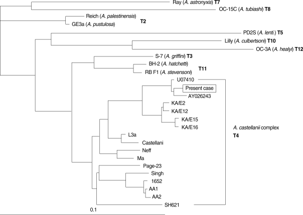
Fig. 3). The causative organism of the case was identified as
A. castellanii complex (T4 genotype) and was genetically closely related to 2 clinical isolates. AY026243 is
A. polyphaga Jones strain, the first
Acanthamoeba keratitis isolate [
33], and the other strain U07410 is also isolated from a keratitis patient in the USA.
Four
Acanthamoeba isolates (KA/E2, KA/E12, KA/E15 and KA/E16) from infected cornea of Korean patients were reported as
Acanthamoeba lugdunensis based on the Mt DNA RFLP and riboprinting of 18s rDNA [
34]. Three isolates, with the exception of KA/E15, revealed the identical Mt DNA RFLPs and riboprint patterns with
Acanthamoeba L3a, the type strain of
A. lugdunensis. Identification of the isolates as
A. lugdunensis was confirmed by 18s rDNA sequence analysis. The sequence difference of the 4 isolates from L3a strain was 0.1% to 0.4%, and that from
A. castellanii Castellani was 1.2% to 1.5%.
Amebic keratitis related to orthokeratology lens overnight wear was reported in a 15-year-old girl who wore the lens for 10 months [
29]. Based on the 18s rDNA sequence analysis, the infected
Acanthamoeba was identified as the genotype T4 and very closely related to
A. castellanii Ma strain which had been isolated also from amoebic keratitis.
Xuan et al. [
35] characterized 3
Acanthamoeba isolates (KA/E9, KA/E17, and KA/E23) from Korean keratitis patients and identified as
Acanthamoeba triangularis by the 18s rDNA sequence homology. The 3 isolates showed 1.3-1.6% of 18s rDNA sequence differences from
A. triangularis. Although the cyst morphology of the 3 isolates was not identical with
A. triangularis, their mitochondrial DNA RFLP was very similar with that of
A. triangularis.
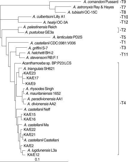
Here we present the phylogenetic tree of
Acanthamoeba by 18s rDNA sequence analysis of 22 known strains as reference strains and 10 clinical isolates (KA/E series) from Korean keratitis patients (
Table 1 &
Fig. 4). All clinical isolates from Korean patients belonged to the morphological group II and identified as the sequence type T4. They scattered into 4 subclades within T4. It is not certain to assign them as separate species or
A. castellanii species complex. By Mt 16S rDNA sequence analysis of Korean keratitis isolates, the strains in the T4 genotype were subgrouped into 5 clades (
Fig. 5).
A. castellanii Castellani and
A. lugdunensis L3a belonged to different clades. With these results, we confirmed the potential of Mt 16s rDNA to identify species in genotype T4 as suggested by Ledee et al. [
31].
Acanthamoeba isolate from Korean soil with strain name KA/S2 was characterized by Chung et al. [
20]. Mt DNA RFLP patterns of KA/S2 were identical with those of the Neff strain originally isolated from the soil of the USA.
By a nationwide survey on the contamination rate of
Acanthamoeba in contact lens storage cases, 110
Acanthamoeba isolates were identified and characterized by comparison of riboprinting and Mt DNA RFLP with those of reference strains obtained from American Type Culture Collections (ATCC) [
36-
38]. All isolates from the lens storage cases belonged to the morphological group II and were identified as the genotype T4. Their individual genetic characteristics by Mt DNA RFLP showed that the most common pattern was that of
A. lugdunensis L3a and KA/E2, the type stain and a corneal isolate from a Korean keratitis patient, respectively. Another common pattern was that of
A. castellanii Castellani and KA/E3, the type strain and another corneal isolate found in Korea. The pattern of
A. castellanii Ma strain, a corneal isolate from the USA was also quite common in some part of Korea. The prevalent pattern for each type of
Acanthamoeba in southwestern area was very different from that in southeastern areas and Seoul, Korea [
38]. Most
Acanthamoeba isolates from domestic tap water were identified as potential keratopathogens based on the molecular characteristics of Mt DNA RFLP and 18s rDNA sequence analysis [
39]. Interestingly, the Mt DNA RFLP patterns of 5 isolates from contact lens storage cases were found to be the same as those of isolates from the lens owner's domestic tap water.
Acanthamoeba isolates from ocean sediments of Korea showed big diversity in genetic characteristics compared to the isolates from contact storage cases [
40]. All of 18 isolates belonged to the morphologic group II. Fifteen isolates showed close relatedness with clinical isolates and
A. castellanii Castellani, and they formed a lineage equivalent to the T4 genotype. Two reference strains from the ocean sediment,
A. hatchetti BH-2 and
A. griffini S-7, clustered unequivocally with these 15 isolates.
Im and Shin reported [
41] an isolate from a freshwater fish in Korea as a new species of
Acanthamoeba and assigned as
Acanthamoeba sohi, based on random amplified polymorphic DNA marker analysis, Mt DNA RFLP, and 18s rDNA sequence analysis [
41]. The isolate belonged to the morphologic group III and formed a different clade from 3 reference strains of group 3,
A. culbertsoni,
A. healyi, and
Acanthamoeba royreba in the phylogenic tree.
CONCLUSION
Although various molecular characteristics have been applied for the taxonomy of Acanthamoeba up to date, hitherto species identification system needs to be improved. Sequence data of 18s rDNA may provide information for genotyping or grouping to the most closely related known species. Mt 16s rDNA sequence analysis could be an additive data to subgroup the strains in the genotype T4, to which most clinical and environmental isolates belong. More detailed molecular characteristics could be obtained by isoenzyme analysis and Mt DNA RFLP. Comparison of molecular characteristics of clinical or environmental isolates with already identified strains is also a short way to identify a new isolate.
ACKNOWLEDGEMENTS
The author thanks Prof. Dong-Il Chung, Kyungpook National University School of Medicine, for careful reading of this manuscript and helpful discussions.
References
- 1. Awwad ST, Petroll WM, McCulley JP, Cavanagh HD. Updates in Acanthamoeba keratitis. Eye Contact Lens 2007;33:1-8.
- 2. Patel DV, McGhee CN. Acanthamoeba keratitis: a comprehensive photographic reference of common and uncommon signs. Clin Experiment Ophthalmol 2009;37:232-238.
- 3. Ringsted J, Jager BV, Suk D, Visvesvara GS. Probable acanthamoeba meningoencephalitis in a Korean child. Am J Clin Pathol 1976;66:723-730.
- 4. Im K, Kim DS. Acanthamoebiasis in Korea: two new cases with clinical cases review. Yonsei Med J 1998;39:478-484.
- 5. Pussard M, Pons R. Morphologie de la paroi kystiqueet taxonomie du genre Acanthamoeba (Protozoa, Amoebida). Protistologica 1977;8:557-598.
- 6. Sawyer T. Acanthamoeba griffini, a new species of marine amoeba. J Protozool 1971;18:650-654.
- 7. Stratford MP, Griffiths AJ. Excystment of the amoeba Hartmannella castellanii. J Gen Microbiol 1971;66:247-249.
- 8. Visvesvara GS. Classification of Acanthamoeba. Rev Infect Dis 1991;13(suppl 5):S369-S372.
- 9. Costas M, Griffiths AJ. The suitability of starch gel electrophoresis of esterases and acid phosphatases for the study of Acanthamoeba taxonomy. Arch Protistenk 1980;123:272-279.
- 10. Costas M, Griffithes AJ. The characterization of 71 strains of Acanthamoeba using starch electrophoresis of isozymes. (Abstr). 7. 4th Int Cong Protozool. Warsaw, Poland: Progress in Protozoology 63.
- 11. Costas M, Griffiths AJ. Enzyme composition and the taxonomy of Acanthamoeba. J Protozool 1985;32:604-607.
- 12. Mathews HM, Moss DM, Healy GR, Visvesvara GS. Polyacrylamide gel electrophoresis of isoenzymes from Entamoeba species. J Clin Microbiol 1983;17:1009-1012.
- 13. De Jonckheere JF. Isoenzyme and total protein analysis by agarose isoelectric focusing, and taxonomy of the genus Acanthamoeba. J Protozool 1983;30:701-706.
- 14. Kong HH, Park JH, Chung DI. Interstrain polymorphisms of isoenzyme profiles and mitochondrial DNA fingerprints among seven strains assigned to Acanthamoeba polyphaga. Korean J Parasitol 1995;33:331-340.
- 15. Bogler SA, Zarley CD, Burianek LL, Fuerst PA, Byers TJ. Interstrain mitochondrial DNA polymorphism detected in Acanthamoeba by restriction endonuclease analysis. Mol Biochem Parasitol 1983;8:145-163.
- 16. Yagita K, Endo T. Restriction enzyme analysis of mitochondrial DNA of Acanthamoeba strains in Japan. J Protozool 1990;37:570-575.
- 17. Gautom RK, Lory S, Seyedirashti S, Bergeron DL, Fritsche TR. Mitochondrial DNA fingerprinting of Acanthamoeba spp. isolated from clinical and environmental sources. J Clin Microbiol 1994;32:1070-1073.
- 18. Byers TJ, Hugo ER, Stewart VJ. Genes of Acanthamoeba: DNA, RNA and protein sequences (a review). J Protozool 1990;37:17S-25S.
- 19. Kilvington S, Beeching JR, White DG. Differentiation of Acanthamoeba strains from infected corneas and the environment by using restriction endonuclease digestion of whole-cell DNA. J Clin Microbiol 1991;29:310-314.
- 20. Chung DI, Kong HH, Yu HS, Oh YM, Yee ST, Lim YJ. Biochemical and molecular characterization of a strain KA/S2 of Acanthamoeba castellanii isolated from Korean soil. Korean J Parasitol 1996;34:79-85.
- 21. Chung DI, Yu HS, Hwang MY, Kim TH, Kim TO, Yun HC, Kong HH. Subgenus classification of Acanthamoeba by riboprinting. Korean J Parasitol 1998;36:69-80.
- 22. Bhattacharya S, Som I, Bhattacharya A. The ribosomal DNA plasmids of Entamoeba. Parasitol Today 1998;14:181-185.
- 23. Khan NA, Paget TA. Molecular tools for speciation and epidemiological studies of Acanthamoeba. Curr Microbiol 2002;44:444-449.
- 24. Kong HH, Chung DI. PCR and RFLP variation of conserved region of small subunit ribosomal DNA among Acanthamoeba isolates assigned to either A. castellanii or A. polyphaga. Korean J Parasitol 1996;34:127-134.
- 25. Gast RJ, Ledee DR, Fuerst PA, Byers TJ. Subgenus systematics of Acanthamoeba: four nuclear 18S rDNA sequence types. J Eukaryot Microbiol 1996;43:498-504.
- 26. Stothard DR, Schroeder-Diedrich JM, Awwad MH, Gast RJ, Ledee DR, Rodriguez-Zaragoza S, Dean CL, Fuerst PA, Byers TJ. The evolutionary history of the genus Acanthamoeba and the identification of eight new 18S rRNA gene sequence types. J Eukaryot Microbiol 1998;45:45-54.
- 27. Schroeder JM, Booton GC, Hay J, Niszl IA, Seal DV, Markus MB, Fuerst PA, Byers TJ. Use of subgenic 18S ribosomal DNA PCR and sequencing for genus and genotype identification of Acanthamoebae from humans with keratitis and from sewage sludge. J Clin Microbiol 2001;39:1903-1911.
- 28. Yu HS, Kong HH, Kim SY, Hahn YH, Hahn TW, Chung DI. Laboratory investigation of Acanthamoeba lugdunensis from patients with keratitis. Invest Ophthalmol Vis Sci 2004;45:1418-1426.
- 29. Lee SJ, Jeong HJ, Lee JE, Lee JS, Xuan YH, Kong HH, Chung DI, Ock MS, Yu HS. Molecular characterization of Acamthamoeba isolated from amebic keratitis related to orthokeratology lens overnight wear. Korean J Parasitol 2006;44:313-320.
- 30. Yu HS, Hwang MY, Kim TO, Yun HC, Kim TH, Kong HH, Chung DI. Phylogenetic relationships among Acanthamoeba spp. based on PCR-RFLP analyses of mitochondrial small subunit rRNA gene. Korean J Parasitol 1999;37:181-188.
- 31. Ledee DR, Booton GC, Awwad MH, Sharma S, Aggarwal RK, Niszl IA, Markus MB, Fuerst PA, Byers TJ. Advantages of using mitochondrial 16S rDNA sequences to classify clinical isolates of Acanthamoeba. Invest Ophthalmol Vis Sci 2003;44:1142-1149.
- 32. Hwang HK, Im KI, Soh CT, Yun DJ. Experimental study on the pathogenicity of free-living amoebae. Yonsei J Med Sci 1976;9:182-194.
- 33. Jones DB, Visvesvara GS, Robinson NM. Acanthamoeba polyphaga keratitis and Acenthamoeba uveitis associated with fatal meningoencephalitis. Trans Ophthalmol Soc (UK) 1975;95:221-232.
- 34. Yu HS, Kong HH, Kim SY, Hahn YH, Hahn TW, Chung DI. Laboratory investigation of Acanthamoeba lugdunensis from patients with keratitis. Invest Ophthalmol Vis Sci 2004;45:1418-1426.
- 35. Xuan YH, Chung BS, Hong YC, Kong HH, Hahn TW, Chung DI. Keratitis by Acanthamoeba triangularis: report of cases and characterization of isolates. Korean J Parasitol 2008;46:157-164.
- 36. Lee SM, Choi YJ, Ryu HW, Kong HH, Chung DI. Species identification and molecular characterization of Acanthamoeba isolated from contact lens paraphernalia. Korean J Ophthalmol 1997;11:39-50.
- 37. Yu HS, Choi KH, Kim HK, Kong HH, Chung DI. Genetic analyses of Acanthamoeba isolates from contact lens storage cases of students in Seoul, Korea. Korean J Parasitol 2001;39:161-170.
- 38. Kong HH, Shin JY, Yu HS, Kim J, Hahn TW, Hahn YH, Chung DI. Mitochondrial DNA restriction fragment length polymorphism (RFLP) and 18S small-subunit ribosomal DNA PCR-RFLP analyses of Acanthamoeba isolated from contact lens storage cases of residents in southwestern Korea. J Clin Microbiol 2002;40:1199-1206.
- 39. Jeong HJ, Lee SJ, Kim JH, Xuan YH, Lee KH, Park SK, Choi SH, Chung DI, Kong HH, Ock MS, Yu HS. Acanthamoeba: keratopathogenicity of isolates from domestic tap water in Korea. Exp Parasitol 2007;117:357-367.
- 40. Liu H, Ha YR, Lee ST, Hong YC, Kong HH, Chung DI. Genetic diversity of Acanthamoeba isolated from ocean sediments. Korean J Parasitol 2006;44:117-125.
- 41. Im KI, Shin HJ. Acanthamoeba sohi, n. sp., a pathogenic Korean isolate YM-4 from a freshwater fish. Korean J Parasitol 2003;41:181-188.
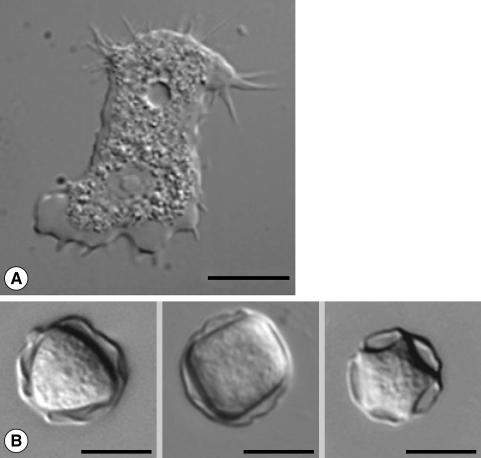
Fig. 1Microphotographs of Acanthamoeba. (A) Trophozoite of A. castellanii Castellani with many characteristic pin-like acanthopodia and numerous vacuoles. Nucleus and a contractile vacuole are prominent. (B) Various shaped cysts of Acanthamoeba with different arm numbers.

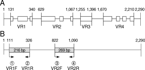
Fig. 2Schematic diagram of 18s rDNA sequence of A. castellanii Casteallani. (A) 4 variable regions of 18s rDNA are demonstrated with white boxes and named as VR1, VR2, VR3, and VR4. (B) Primer sets for PCR of 2 parts of variable regions are presented.

Fig. 3Phylogenetic dendrogram derived by aligning the partial 18s rDNA from the present case and various strains of Acanthamoeba in the database. U07410 and AY026243 are Genbank accession numbers of 18s rDNA sequences of the 2 Acanthamoeba isolates.

Fig. 4Neighbour-joining dendrogram based on comparative 18s rDNA sequence analysis showing relationships of Acanthamoeba isolates from Korean keratitis patients to representative members of the genus Acanthamoeba.

Fig. 5Phylogenetic analysis using ClustalX and TreeView for Mt 16s rDNA sequence analysis showing relationships of Acanthamoeba isolates from Korean keratitis patients to representative members of the genus Acanthamoeba. T1-T12 mean the sequence type of the strains by 18s rDNA sequence analysis.
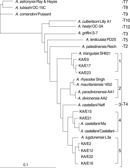
Table 1.
Acanthamoeba strains used in the present study for reference strains and clinical isolates
Table 1.
|
No |
Species and strain |
ATCC No. |
18s rDNA Accession No. |
Source |
Geographic source |
|
1 |
A. astronyxis Ray & Hayes |
30137 |
AF019064 |
Lab water |
USA |
|
2 |
A. tubiashi OC-15C |
30867 |
AF019065 |
Fresh water |
USA |
|
3 |
A. comandoni Pussard |
30135 |
AF019066 |
Soil |
France |
|
4 |
A. lenticulata PD2S |
30841 |
U94741 |
swimming pool |
France |
|
5 |
A. culbertsoni Lilly A1 |
30171 |
AF019067 |
human cell culture |
USA |
|
6 |
A. healyi OC-3A |
30866 |
AF019070 |
GAE |
BWI |
|
7 |
A. palestinensis Reich |
30870 |
U07411 |
Soil |
Israel |
|
8 |
A. griffini S-7 |
30731 |
U07412 |
Beach bottom |
USA |
|
9 |
A. castellanii Neff |
50373 |
U07416 |
Soil |
USA |
|
10 |
A. castellanii Castellani |
50374 |
U07413 |
Yeast culture |
UK |
|
11 |
A. castellanii Ma |
50370 |
U07414 |
Keratitis |
USA |
|
12 |
A. rhysodes Singh |
30973 |
AY351644 |
Soil |
UK |
|
13 |
A. mauritaniensis 1652 |
50253 |
AY351647 |
Soil |
UK |
|
14 |
A. paradivionensis AA1 |
50251 |
AY351645 |
Soil |
France |
|
15 |
A. divionensis AA2 |
20238 |
AY351646 |
Soil |
Morocco |
|
16 |
A. triangulais SH621 |
50254 |
AF316547 |
human feces |
France |
|
17 |
A. pustulosa GE3a |
50252 |
AF019050 |
soil |
USA |
|
18 |
A. castellanii CDC:0981:V006 |
- |
U07400 |
|
|
|
19 |
A. lugdunensis L3a |
50240 |
AF005995 |
fresh water |
France |
|
20 |
Acanthamoeba sp. BP:P23:LCS |
- |
FJ422546 |
|
|
|
21 |
A. stevensoni RB:F:1 |
50388 |
AF019069 |
|
|
|
22 |
A. hatchetti BH-2 |
30730 |
AF019068 |
sediment |
USA |
|
23 |
Acanthamoeba sp. KA/E2 |
- |
AF005998 |
|
|
|
24 |
Acanthamoeba sp. KA/E9 |
- |
AF316546 |
keratitis |
South Korea |
|
25 |
Acanthamoeba sp. KA/E12 |
- |
AF316545 |
keratitis |
South Korea |
|
26 |
Acanthamoeba sp. KA/E15 |
- |
AY148961 |
keratitis |
South Korea |
|
27 |
Acanthamoeba sp. KA/E16 |
- |
AY148962 |
keratitis |
South Korea |
|
28 |
Acanthamoeba sp. KA/E17 |
- |
AY148963 |
keratitis |
South Korea |
|
29 |
Acanthamoeba sp. KA/E21 |
- |
EF140633 |
keratitis |
South Korea |
|
30 |
Acanthamoeba sp. KA/E22 |
- |
EF140638 |
keratitis |
South Korea |
|
31 |
Acanthamoeba sp. KA/E23 |
- |
EF140625 |
keratitis |
South Korea |