Abstract
In November 2007, a 46-year-old male Thai patient presented with chronic abdominal pain for over 3 years. Colonoscopy revealed a small parasite of about 2 × 1 mm in size attached to the cecum mucosa. The worm was removed endoscopically, fixed, and stained for morphological observations. The specimen was identified as Anchitrema sanguineum (Digenea: Anchitrematidae), a trematode first reported in a reptile, Chamaeleo vulgaris, from Egypt, and then sporadically found in the intestines of insectivorous bats and other mammals. The patient was treated with praziquantel but no more worms were found in his stool. His symptoms improved slightly but not cured completely. It remains unclear whether the chronic abdominal pain of the patient was caused by this trematode infection. Whatever is the pathogenicity of this trematode, this is the first human case of A. sanguineum infection in the literature.
-
Key words: Anchitrema sanguineum, human, colonoscopy, Thailand
INTRODUCTION
Anchitrema sanguineum is a rare trematode parasite first found in the gut of a reptile,
Chamaeleo vulgaris, in Egypt and named
Distomum sanguineum Sonsino, 1894. Later this parasite was found in insectivorous bats in Egypt and renamed as
A. sanguineum [
1]. Subsequently, this trematode was sporadically found in the intestines of insectivorous bats in India [
2], Malaysia [
3], Thailand [
4], Taiwan [
5], and Japan [
6]. A massive infection of
A. sanguineum in the stomach and intestines of cameleons was reported in Hyderabad, India [
7]. Apart from bats and cameleons, this parasite was found in rats in Egypt [
8] and Thailand [
9]. The morphology of adult worms was re-described in detail [
10]. However, little is known about the life cycle or other biological features of
A. sanguineum, and human infection has never been reported. Here, we report the first case of
A. sanguineum found in the human colon in Thailand.
CASE RECORD
In November 2007, a 46-year-old male Thai resident in Bangkok visited Phayathai Hospital, a private hospital in Bangkok, with a history of chronic abdominal pain and discomfort since January 2004. He had visited several hospitals before and received some medication, but the pain did not improve. Apart from the abdominal pain, he had no history of chronic diseases.
On his visit, he had mild lower abdominal pain with tenderness down to the perineum. The pain referred to both legs, but with no neurological deficits. Physical examinations showed normal blood pressure (113/73 mm/Hg), no weight loss, no pallor, no jaundice, and no abnormal neurological signs. The complete blood count and liver function test results were normal.
Colonoscopic observation showed a small parasite (2 × 1 mm) attached to the cecum mucosa without any apparent damage of the mucosa (
Fig. 1). The worm was removed, fixed in 10% formalin, stained with acetic carmine, and mounted in Permount® for morphological identification. After removal of the worm by endoscopy, the patient was treated with 25 mg/kg (3 times per day) oral dose of praziquantel, but no more worms were expelled. His symptoms improved slightly, but without cure.
Since the parasite was identified as A. sanguineum, we asked the patient about his history of possible contact with bats, reptiles, or other animals. Ten years beforehand, he had worked at Jompol Cave, Ratchaburi province, Thailand, for a few days. The cave was a resting area for bats. Since then, he never visited Jompol Cave again. Recently he has been living in Bangkok with a pet guinea pig which he bought from Jatujak market in Bangkok 2 years ago. Although we examined the stools of his pet guineapig, they were negative for trematode or any other parasite eggs.
IDENTIFICATION OF THE WORM
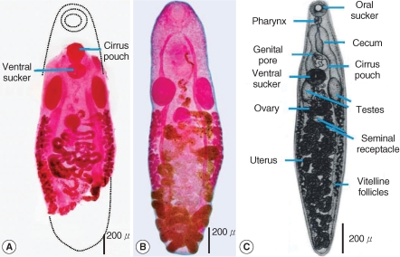
The anterior tip and the posterior 1/3 of the parasite were broken and missing during endoscopic extirpation and further processing. Nevertheless, morphologically informative parts of the body were preserved well as shown in
Fig. 2A. The oral sucker and the pharynx were missing. The intestinal furca were also missing down to the shoulders. The intestinal ceca pass laterally towards the end of the body. The ventral sucker was round and about 200 µm in diameter. The cirrus pouch was almost round in shape and lies anterior to the ventral sucker. Two elongated oval testes measuring 400 × 160 µm (right) and 380 × 150 µm (left) were located postero-laterally to the ventral sucker. A single ovary lied in the middle part of the body. The vitelline follicles were scattered bilaterally in the mid-body. The uterus twisted irregularly and occupied the posterior part of the body. The intra-uterine eggs were small, about 22.93 (20.0-25.0) µm long and 11.80 (10.0-15.0) µm wide, and morphologically similar to opisthorchid eggs. Those morphological features were compared with the in-house specimen of
A. sanguineum recovered from the intestine of a puppy in Bangkok (
Fig. 2B) and the schematic drawing of
A. sanguineum found in the intestine of a rat in Thailand (
Fig. 2C; ref. [
9] with permission). Moreover, morphometric data of those materials were compared further with those described for Egyptian samples (
Table 1). Apparently from those morphological and morphometric observations, the specimen obtained from the patient could be identified as
A. sanguineum.
DISCUSSION
Anchitrema is a relatively small genus, with 5 nominal species recognized as valid species by Yamaguti [
11];
A. sanguineum (Sonsino, 1894) Looss, 1899,
A. latum Gedoelst, 1919,
A. sokolowi (Skrjabin, 1914) Dollfus, 1929,
A. philippinorum (Tubangui, 1928) Yamaguti, 1958, and
A. congolense (Sandground, 1937) Yamaguti, 1958. Two more species were proposed from India;
A. indicum (Thaper, 1931) Pande, 1935 and
A. lucknowensis Agrawal, 1966. However, after extensive morphological study by Saoud and Ramadan [
10],
A. congolense was found to be a synonym of
A. latum, and
A. lucknowensis a synonym of
A. philippinorum. Instead, they added
A. longiformis as a new species [
10]. Therefore, a total of 6 species are now recognized as valid species in the genus
Anchitrema, all of which are primarily parasites of insectivorous bats.
A. sanguineum is widely distributed in the tropical to subtropical areas from Egypt to southern Japan. In Thailand,
A. sanguineum was first found in bats in Nongkhai province [
4], and then in rats in Kanchanaburi province [
9]. The present results show that a dog and a human can act as accidental hosts for
A. sanguineum adult worms, suggesting a low host specificity of this parasite.
Until now, the precise life cycle of this parasite remains unknown. However, based on its morphological similarities and the range of mammalian hosts,
A. sanguineum is supposed to be similar to
Dicrocoelium dendriticum, which accidentally infect humans and sometime cause abdominal symptoms [
12]. Humans might be accidental hosts for
A. sanguinarum, and ingestion may occur via infected insects or contaminated food or water. Usually small fluke infections are asymptomatic or just causing vague abdominal discomfort. In the present study, abdominal pain of the patient persisted after the removal of the worm by endoscopy and praziquantel treatment. Moreover, the clinical manifestation did not correlate with the site of the parasite attachment. Therefore, this patient's clinical condition was not solely attributable to this trematode infection.
In conclusion, this is the first record of a human infection with A. sanguineum. To elucidate the transmission route of this trematode, potential reservoir hosts should be studied further.
ACKNOWLEDGEMENTS
The authors would like to thank the patient for agreeing to reveal more details and also thankful to Prof. Dr. Yukifumi Nawa and Paul Adams for editing the manuscript.
References
Fig. 1Colonoscopic observation showing a small parasite (1 × 2 mm) attached to the cecum mucosa.
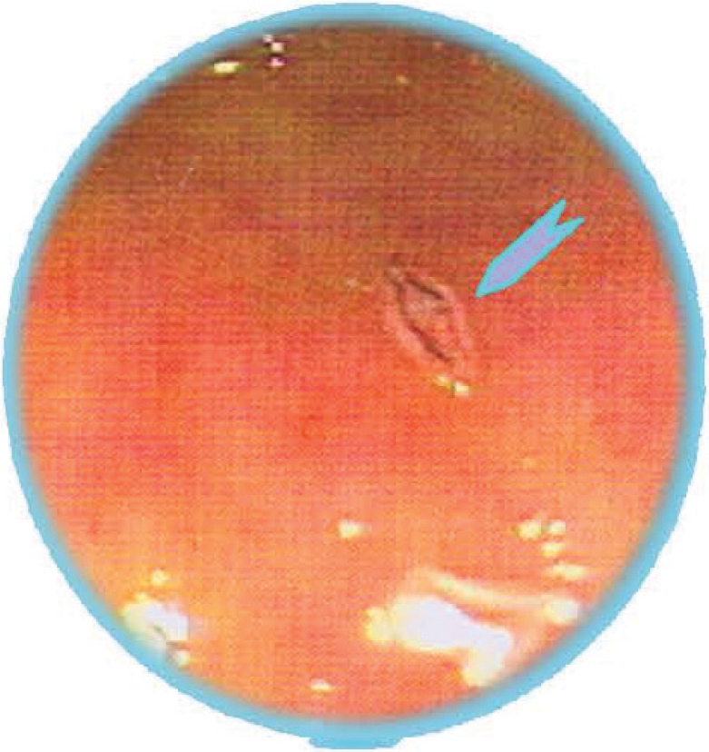
Fig. 2
Anchitrema sanguineum adults. (A) The worm from colonoscopy of the present case. (B) An in-house specimen from the intestine of a puppy in Bangkok. (C) Diagram of a worm recovered from
Rattus rattus [
9].

Table 1.Morphometric comparison of Anchitrema sanguineum from the patient and other sources
Table 1.
|
From the patient |
In house specimena
|
From a rat of Thailandb
|
From bats of Egyptc
|
|
Body (L × W) |
? × 0.87 |
2.08 × 0.96 |
NDd
|
1.79-3.73 × 0.63-0.98 |
|
Oral sucker |
Missing |
220 × 260 |
220 × 253 |
230-250 × 210-320 |
|
Ventral sucker |
200 |
170 × 180 |
205 |
180-320 × 180-280 |
|
O/V ratio |
ND |
1.37 |
1.15 |
1-1.4 : 1 |
|
Cirrus pouch |
200 × 220 |
0.18 × 0.19 |
ND |
140-260 × 130-230 |
|
Testis (right) |
400 × 160 |
260 × 230 |
418 × 213 |
320-490 × 180-250 |
|
Testis (left) |
380 × 150 |
270 × 180 |
319 × 187 |
320-480 × 180-250 |
|
Ovary |
170 |
180 |
176 |
180-240 × 150-210 |
|
Eggs |
22.93 × 11.8 |
26.88 × 14.50 |
25.6 × 14.8 |
19-23 × 9-10 |