Abstract
A 3-yr-old female mongrel dog was referred to the Veterinary Teaching Hospital of Chungnam National University in the Republic of Korea. An adult heartworm, Dirofilaria immitis, was found in the abdominal cavity of the dog during spaying. Dirofilariasis in this dog was also diagnosed by modified Knott's test, ELISA test, and PCR analysis. The present case is the first report on the migration of an adult dog heartworm to the abdominal cavity of a dog in the Republic of Korea.
-
Key words: Dirofilaria immitis, dog, migration, abdominal cavity
INTRODUCTION
The heartworm disease is an infectious disease of dogs with
Dirofilaria immitis combined with cardiovascular and circulatory abnormalities. The heartworm disease can become a serious health risk when associated with a severe infection. Adult heartworms normally live in the pulmonary artery and right ventricle [
1]. Much less frequently, immature 5th-stage larvae (L
5) may migrate aberrantly to other sites, including the brain, spinal cord, liver, epidural space, anterior chamber of the eye, vitreous, and peritoneal cavity [
1]. In Republic of Korea, Pak and Lee [
2] first reported that microfilaremic rate of the canine heartworm was 21% in Chinju area, and Song et al. [
3] reported that 339 (40%) of 848 samples tested with antigen detecting ELISA kits showed positive reactions for
D. immitis in 4 different provinces. The present case is the first report on migration of an adult heartworm to the abdominal cavity of a dog nonetheless the Republic of Korea is an enzootic area with a high prevalence of dirofilariasis.
CASE REPORT
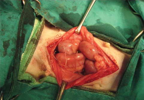
A 3-yr-old female mongrel dog for spaying was referred to the Veterinary Teaching Hospital of Chungnam National University, Republic of Korea. The physical examination showed no abnormal clinical signs or findings. The complete blood count and serum chemistry profiles were within normal ranges, except for aspartate transaminase 161 U/L (normal reference range; 22-84) and, creatine kinase 1,400 U/L (normal reference range; 51-485). A long slender nematode was found in the abdominal cavity of the dog during the spaying (
Fig. 1). This parasite was a 12 cm long male heartworm with a coiled tail portion. Many microfilariae were detected by a direct blood smear and Modified Knott's test [
4]. Electrocardiography showed a deep S wave pattern, which indicates a right ventricle hypertrophy. This was attributed to an infection of many adult worms in the right ventricle and pulmonary artery. A canine heartworm antigen detecting kit (SNAP test, IDEXX Laboratories, Westbrook, Maine, USA) was employed according to the protocol provided by the manufacturer. The ELISA kit test showed a strong positive reaction, which was attributed to a heavy infection of
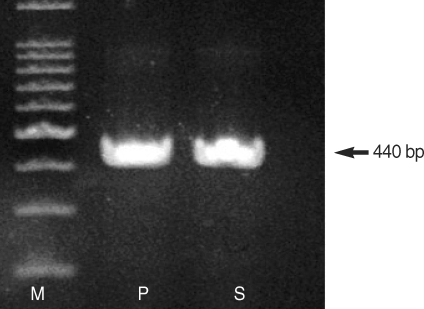
D. immitis. The worm found in the abdominal cavity was lysed in 0.1 M Tris-HCl (pH 8.0) containing 1% SDS, 0.1 M NaCl, and 10 mM EDTA. The samples were treated with proteinase K (100 µ/ml) at 55℃ for 2 hr. The DNA was extracted with phenol/chloroform, precipitated by ethanol, and then dissolved in 50 µl of a TE buffer (10 mM Tris-HCl pH 8.0, 1 mM EDTA). The primers used for PCR amplification were 5'-GCATCTTAGAACTTGGT CCATCC-3'(forward), and 5'-CAAGGCGTATTTACCGCCGAC-3'(reverse), which have been described by Watts et al. [
5] for the amplification of the 440-bp 16S rRNA gene segment. PCR was performed in 50 µl reaction mixtures, which contained 5 µl template DNA, 2 µl 20 pmol of each primer, 4 µl 200 mM of each dNTP, 1.25 U rTaq DNA polymerase (Takara, Tokyo, Japan), and 5 µl 10 × PCR buffer (10 mM Tris-HCl, 1.5 mM MgCl
2, and 0.001% gelatin). The thermal cycler was set at 94℃ for 3 min, to activate the polymerase, which was followed by 30 cycles of denaturation at 94℃ for 30 sec, annealing at 55℃ for 40 sec and extension at 72℃ for 50 sec. A 5-µl sample of the PCR products was then analyzed by electrophoresis in 0.8%-agarose gel followed by ethidium-bromide staining and photography. PCR analysis for an adult worm revealed a heartworm DNA-positive reaction (
Fig. 2). Therefore, it was also confirmed that the worm detected in the abdominal cavity was
D. immitis by PCR analysis.
DISCUSSION
The Republic of Korea is one of the enzootic regions of
D. immitis, and the prevalence in dogs appears to have increased recently [
3,
6]. This heartworm is transmitted by many different species of mosquitoes [
1]. Lee et al. [
7] reported the detection of heartworm DNA in mosquitoes, such as
Anopheles sinensis,
Aedes vexans nipponii and
Culex pipiens in Gyeonggi-do and Gangwon-do, the Republic of Korea. Dogs with aberrantly located adult heartworms have been reported since 1856, and Tada et al. [
8] reported a human case of
D. immitis infection occurring in the abdominal cavity. Heartworms have been found in many body sites; however, the mechanism for the aberrant location of heartworms has not been determined [
9]. Dibbell [
10] first reported the detection of a female
D. immitis in the abdominal cavity, but no microfilariae were observed in the blood. Often there is no evidence of heartworms present, and the aberrant worm is not discharging microfilariae. The simultaneous occurrence of worms in their normal habitat causes parasitemia [
11]. In our case, many microfilariae were observed by optical microscopy, which may be due to the concurrent infection of
D. immitis in the right ventricle and pulmonary artery. Treatment of aberrantly migrating heartworms requires either nothing, surgical excision of the offending parasite, adulticidal therapy, or symptomatic treatment [
1]. Removal of the heartworm from the abdominal cavity is recommended if it affects the gastrointestinal tract, indirectly. Further studies are needed to elucidate the mechanism for the ectopic migration of heartworms.
References
Fig. 1An adult Dirofilaria immitis was found in the abdominal cavity of a dog during spaying.

Fig. 2PCR-based detection of D. immitis DNA in an adult worm found in the abdominal cavity of the dog. M is a 100-bp marker. P is a D. immitis DNA-positive control. S is an adult worm collected from abdominal cavity.

Citations
Citations to this article as recorded by

- Subcutaneous dirofilariasis due to Dirofilaria immitis in a dog in Brazil: first report
Welitânia Inácia da Silva, Alexander Rodrigo Dantas Gomes, Maria Carolina de Francisco, Janete Madalena da Silva, Hodias Sousa de Oliveira Filho, Thais Ferreira Feitosa, Vinícius Longo Ribeiro Vilela
Revista Brasileira de Parasitologia Veterinária.2023;[Epub] CrossRef - Two cases of ectopic dirofilariasis by Dirofilaria immitis in subconjunctival and subcutaneous tissues in dogs
Yeong-Seok Goh, Hye-Min Kim, Badriah Alkathiri, Hong Suh Chang, Young Min Yoon, Seung-Hun Lee, Kyung-Mee Park
Parasitology International.2023; 92: 102683. CrossRef - Incidental Finding of Dirofilaria immitis (Spirurida: Onchocercidae) Microfilariae in the Bone Marrow of a Dog with Mixed Leishmania infantum-Dirofilaria immitis Infection
Ilaria Lensi, George Lubas, Roberto Amerigo Papini
Zoonotic Diseases.2023; 3(2): 162. CrossRef - Distribution of dirofilariasis in Omsk region
T. S. Ryazanova, A. V. Sverdlova, O. Yu. Starostina, A. A. Nikitin, N. Yu. Grigorova, Yu. V. Kochetkov
Acta Biomedica Scientifica.2022; 7(3): 277. CrossRef - Prevalence of Dirofilaria immitis in shelter dogs in Bucaramanga metropolitan area, Colombia
Angel Alberto Florez Muñoz, Ariel Rosas Martinez, Juan Carlos Pinilla
Veterinary Parasitology: Regional Studies and Reports.2020; 22: 100489. CrossRef - Dracunculus infections in domestic dogs and cats in North America; an under-recognized parasite?
Brianna M. Williams, Christopher A. Cleveland, Guilherme G. Verocai, Liandrie Swanepoel, Kevin D. Niedringhaus, Kelsey L. Paras, Yoko Nagamori, Susan E. Little, Andrea Varela-Stokes, Nicole Nemeth, Heidi Wyrosdick, Alison Tucker, Leigh Deal, Dawn Gauthier
Veterinary Parasitology: Regional Studies and Reports.2018; 13: 148. CrossRef - Aberrant peritoneal localization of Dirofilaria repens in a dog
Marco Pierantozzi, Giada Di Giulio, Donato Traversa, Giovanni Aste, Angela Di Cesare
Veterinary Parasitology: Regional Studies and Reports.2017; 10: 62. CrossRef - A combined human case of Dirofilaria ursi infection in dorsal subcutaneous tissue and Anisakis simplex sensu stricto (s.s.) infection in ventral subcutaneous tissue
Minoru Yamada, Namiko Shishito, Yoshihiro Nozawa, Shigehiko Uni, Keisuke Nishioka, Takaaki Nakaya
Tropical Medicine and Health.2017;[Epub] CrossRef - Aberrant migration of Dirofilaria immitis in the peritoneum of a dog
Courtenay Brines, Prince Agbedanu, Rachel Juelsgaard, Steve Carlson, Matthew Brewer
Veterinary Record Case Reports.2016;[Epub] CrossRef - Bilateral femoral arterial dirofilariasis caused by Dirofilaria immitis in a dog
David A Upchurch, Daniel M Ogden, David G Baker
Veterinary Record Case Reports.2015;[Epub] CrossRef - Retrospective study of canine heartworm disease with caval syndrome in Grenada, West Indies
A. Chikweto, M.I. Bhaiyat, M. Lanza-Perea, S. Veytsman, K. Tiwari, C. De Allie, R.N. Sharma
Veterinary Parasitology.2014; 205(3-4): 721. CrossRef - Vector-borne helminths of dogs and humans in Europe
Domenico Otranto, Filipe Dantas-Torres, Emanuele Brianti, Donato Traversa, Dusan Petrić, Claudio Genchi, Gioia Capelli
Parasites & Vectors.2013;[Epub] CrossRef - Ectopic dirofilariosis in two dogs from Rio de Janeiro state, Brazil
Beatriz Brener, Patricia Riddell Millar, Danuza Pinheiro Bastos Garcia de Mattos, Flávia Uchôa, Bethânia Bastos, Ingrid Rodrigues Lyrio, Pedro Luis Aragon, Adriana Pittella Sudré
Revista do Instituto de Medicina Tropical de São Paulo.2012; 54(3): 175. CrossRef