Abstract
Accurate identification of tick species is essential for understanding their role in pathogen transmission and guiding vector control strategies. Although morphological classification is widely used, it can be limited by specimen damage or immature developmental stages. In this study, we comparatively evaluated mitochondrial 16S and 12S ribosomal DNA (rDNA) markers for species-level identification and morphological verification of hard ticks and rodent-associated ectoparasites collected from vegetation, wild rodents, and patients in Gwangju, Republic of Korea. A total of 74 morphologically identified adult ticks—Haemaphysalis longicornis, Haemaphysalis flava, and Ixodes nipponensis—along with 3 damaged ticks removed from patients, were analyzed. PCR and sequencing success varied among species and markers. The 16S rDNA marker showed a higher amplification rate but lower sequencing success, whereas 12S rDNA showed more consistent performance. I. nipponensis showed the most balanced performance (89.5% for 16S and 78.9% for 12S), H. flava showed moderate variation, and H. longicornis demonstrated strong marker dependence, with low 16S but complete 12S success. Importantly, 16S rDNA identified 3 severely damaged ticks from patients—2 I. nipponensis and 1 Amblyomma testudinarium—demonstrating its diagnostic value when morphology was uninformative. Phylogenetic analyses of both markers yielded well-supported clades consistent with morphological identification. Additionally, 12S rDNA identified 3 flea specimens (Ctenophthalmus sp. and Neopsylla specialis) obtained during tick collection from wild rodents, indicating exploratory applicability to other rodent-associated ectoparasites. These findings support mitochondrial 16S and 12S rDNA markers as reliable tools for tick identification, especially when morphological assessment is limited by damage or developmental stage.
-
Key words: Ticks, Haemaphysalis flava, Haemaphysalis longicornis, Ixodes nipponensis, mitochondrial DNA, polymerase chain reaction
Introduction
Vector-borne infectious diseases transmitted by mosquitoes, ticks, fleas, and other arthropod vectors account for over 17% of all infectious diseases worldwide [
1]. In Korea, 5 genera and 27 species of ixodid ticks have been identified [
2]. Among these, hard ticks play a critical role as vectors for various pathogens, including viruses, rickettsiae, and protozoa, which affect both humans and animals [
3-
5]. Interest in tick-borne diseases has increased significantly since
Haemaphysalis longicornis, one of the most widespread tick species in East Asia, was identified as the primary vector of severe fever with thrombocytopenia syndrome virus in China, Japan, and Korea [
6,
7].
Accurate species-level identification of ticks is essential for understanding their role in pathogen transmission and developing effective control strategies [
8,
9]. However, morphological identification is often hampered by intraspecific variation, cryptic species, specimen damage during collection or detachment from hosts, and the presence of immature developmental stages [
10]. These limitations become particularly pronounced when closely related species exhibit similar morphological features, making it difficult to differentiate based solely on external traits. In such cases, molecular identification using DNA extracted from tick specimens provides a reliable and precise alternative for taxonomic classification [
11].
Molecular approaches have proven useful in addressing species-level identification challenges in tick taxonomy. For example, in China, genetic analysis using partial 16S ribosomal DNA (rDNA) and the second internal transcribed spacer (ITS-2) sequences successfully distinguished
H. longicornis from the morphologically similar
Haemaphysalis qinghaiensis Teng, 1980, demonstrating clear interspecific divergence and supporting the utility of molecular tools in differentiating closely related tick species [
12]. In addition, Lv et al. [
13] reported the value of several genetic markers, including cytochrome oxidase I (COI), 16S rDNA, ITS-2, and 12S rDNA, for identifying hard tick species. Among these, mitochondrial 16S and 12S rDNA regions are particularly useful because of their species-specific variability and moderate evolutionary rates, making them suitable for identifying damaged, engorged, or immature specimens that are difficult to classify morphologically [
12,
14].
In Korea, direct comparisons between morphological identification and mitochondrial 16S and 12S rDNA-based molecular identification remain limited for ticks collected from diverse sources, particularly for damaged specimens removed from patients. In this study, hard ticks were collected from vegetation, wild rodents, and patients in Gwangju, Korea, and additional rodent-associated ectoparasites obtained during tick collection from wild rodents were included to evaluate the broader applicability of this molecular approach. Morphological identification was compared with molecular identification based on partial 16S and 12S rDNA sequences. Accordingly, this study aimed to compare morphological identification with mitochondrial 16S and 12S rDNA-based molecular identification in ticks collected from vegetation and wild rodents, as well as damaged tick specimens removed from patients.
Methods
Ethics statement
This study was approved by the Ethics in Human Research Committee of Chosun University Hospital under an institutional review board, which approved all the experiments that used ticks removed from tick-bitten humans (approval No. CHOSUN NON2019-001). The institutional review board approved the protocol with verbal consent for the use of ticks and not human participants. We read and explained the verbal version of the consent form to use samples for research purposes and obtained individual consent.
This study was also approved by the Institutional Animal Care and Use Committee of Chosun University. All animals were used in accordance with the approved animal use protocol issued by the Institutional Animal Care and Use Committee of Chosun University for the capture, handling, and sampling of wild rodents.
Tick collection
Tick collection from vegetation by flagging and dragging
From April to November 2014, hard ticks were collected from 5 forested and park-adjacent sites near urban areas in Gwangju, Korea, using standard flagging and dragging techniques. A piece of flannel was swept across the vegetation and any ticks attached to the fabric were carefully detached using forceps. Each specimen was placed in a labeled vial containing information on the collection site, date, and time. All collected specimens were preserved for subsequent morphological and molecular analyses.
Tick collection from wild rodents using Sherman live traps
In October and November 2014, to collect ticks parasitizing wild rodents, Sherman live traps were deployed in 2 forested areas of Gwangju [
15]. Biscuit bait coated with peanut butter was used as the attractant. During the summer months, traps were set at dusk and retrieved the next morning; during winter, to prevent rodent hypothermia, traps were set at 16:00 and collected at 22:00 on the same day. After capture, the rodents were anesthetized in accordance with an approved animal use protocol, and biological samples, such as blood and internal organs, were harvested for parallel studies. Ectoparasitic ticks were collected by suspending rodents head-down above water-filled containers and allowing the detached ticks to fall naturally. Tick specimens obtained from rodents were processed following the same procedures as vegetation-derived samples.
Tick collection from tick-bitten patients
Between January 2015 and December 2018, 3 partially damaged ticks that had been removed from 3 patients after tick bites were collected at Chosun University Hospital, Korea. These patient-derived specimens were handled and examined using the same identification and washing protocols described in the ‘Imaging and washing procedures’ section.
Tick processing, imaging, and DNA extraction
Imaging and washing procedures
Only adult ticks collected using the aforementioned methods were selected for morphological examination. Morphological identification was conducted under a stereomicroscope using standard taxonomic keys [
16], and both dorsal and ventral surfaces of each specimen were photographed. Prior to DNA extraction, ticks were surface-sterilized by immersion in a 10% diluted bleaching solution containing 3%–8% sodium hypochlorite and 0.01%–0.05% sodium hydroxide for 1–2 min. This was followed by sequential rinsing with 70% ethanol (2–5 min) and sterile distilled water (3 min). Individual ticks (~3 mm in size) were then transferred to the wells of sterile U-bottom plates, and the washing solutions were replaced at each step.
Homogenization and DNA extraction
Washed ticks were dried on sterile absorbent paper and transferred into grinding tubes prefilled with 600 μl of lysis buffer and a 2.8-mm stainless steel bead. The buffer consisted of sterile phosphate-buffered saline supplemented with 10% fetal bovine serum and 5% penicillin-streptomycin. Homogenization was performed twice using a FastPrep-24 Classic instrument (MP Biomedicals) at 6,000 rpm for 25 sec. Genomic DNA was extracted from the homogenate using a QIAamp Tissue & Blood Mini Kit (Qiagen), following the manufacturer’s protocol.
PCR amplification of 16S and 12S rDNA
Conventional PCR was performed to amplify the mitochondrial 16S and 12S rDNA fragments for species identification of ticks. For 16S rDNA amplification, the primer pair 16s+1F and 16S-1R was used, and for 12S rDNA amplification, the primer set T1B and T2A was used [
12,
14] (
Table 1).
The 16S rDNA region was amplified using AmpliTaq Gold 360 Master Mix (Applied Biosystems) in a total volume of 20 μl. PCR cycling conditions were as follows: initial denaturation at 95°C for 5 min; 40 cycles of 95°C for 45 sec, 55°C for 1 min, and 72°C for 90 sec; followed by a final extension at 72°C for 5 min. PCR products were electrophoresed on a 1.2% agarose gel and visualized by ethidium bromide staining. Amplification of the 12S rDNA fragment was carried out using AccuPower PCR PreMix (Bioneer) in a 20 μl reaction volume. The PCR condition included an initial denaturation at 95°C for 5 min, followed by 30 cycles of 95°C for 15 sec and 53°C for 45 sec, with a final extension step at 72°C for 5 min. The PCR was performed using an AB thermal cycler (Applied Biosystems). Amplified products were similarly run on a 1.2% agarose gel and stained with ethidium bromide for visualization. The primer sequences are listed in
Table 1.
PCR amplicons of 16S and 12S rDNA were excised from the agarose gels and purified using a QIAquick Gel Extraction Kit (Qiagen). Bidirectional sequencing was performed using specific PCR primers (Solgent). The resulting sequences were searched against the GenBank database using the Basic Local Alignment Search Tool (BLAST) available through the National Center for Biotechnology Information (NCBI), and sequence assembly and alignment were performed using Lasergene software version 8 (DNASTAR). Multiple sequence alignment was performed using ClustalX, the Windows-based interface of Clustal W, with default parameters. Phylogenetic analysis was conducted using the neighbor-joining method based on genetic distances. The robustness of the inferred tree was assessed by bootstrap analysis with 1,000 replicates using the Kimura 2-parameter model. This approach was used to evaluate species-level clustering and concordance with morphological identification based on partial mitochondrial 16S and 12S rDNA sequences.
Results
Morphological identification of tick species
H. longicornis and Haemaphysalis flava ticks were collected by flagging and dragging from forested areas, whereas Ixodes nipponensis ticks were collected from wild rodents captured using Sherman live traps. Only adult ticks were selected for the morphological analysis.
The most frequently collected species was
H. longicornis, comprising 30 adult specimens (No. 60–89), all of which were female; no males were detected. Among the 24
H. flava specimens (No. 1–24), 13 were male and 11 were female. All 20
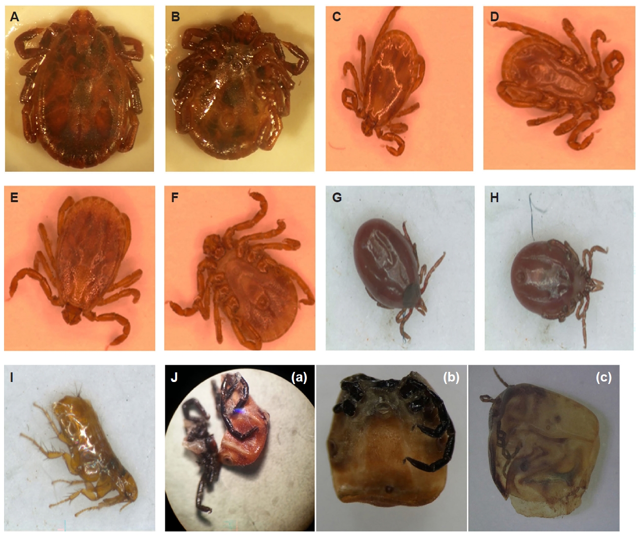
I. nipponensis (No. 30–49) collected from wild rodents were female (
Fig. 1A-
J). In addition to ticks, various ectoparasites such as suspected fleas (No. 25–29), lice (No. 50–54), and mites (No. 55–59) were recovered from rodents.
In addition to the ticks collected from vegetation and rodents, 3 ticks were obtained from patients who had experienced recent tick bites. These specimens were partially damaged during removal, and could not be readily identified by morphological examination. Among them, 2 were tentatively classified under stereomicroscopy as
I. nipponensis (
Fig. 1J) and
Amblyomma testudinarium (
Fig. 1J), respectively; however, owing to specimen conditions, further confirmation via molecular analysis was deemed necessary. The third tick was too extensively damaged for morphological evaluation (
Fig. 1Ja), and was directly subjected to molecular identification.
Molecular identification based on 16S rDNA sequence analysis revealed species-dependent variation in the PCR amplification and sequencing success for molecular identification. Of the 74 morphologically identified tick specimens, 73 (98.6%) produced positive PCR amplicons. Sequencing was performed for all 73 PCR-positive samples, resulting in 38 (52.1%) high-quality sequences suitable for species-level identification (
Table 2). All obtained sequences were verified using BLAST analysis against the NCBI nucleotide database.
H. flava sample (No. 1–24) exhibited a 100% PCR amplification rate (24/24), with 13 of 24 samples (54.2%) yielding sequence data suitable for molecular identification. The 16S rDNA sequences from 3 H. flava specimens (No. 11, 12, and 18) showed 98.6%–99.4% identity with the H. flava mitochondrial genome (GenBank accession No. NC_005292.1), originally isolated in Japan. The remaining ten 16S rDNA sequences showed 98.9%–100% identity with the H. flava isolate H. flava/HA2-16 large subunit ribosomal RNA gene, partial sequence (GenBank accession No. PP346283.1), which was originally isolated in China.
H. longicornis (No. 60–89) also showed 100% PCR amplification (30/30), but only 8 of 30 samples (26.7%) yielded sequence data suitable for molecular identification. These sequences shared 99.2%–99.7% identity with the H. longicornis mitochondrial genome (GenBank accession No. NC_037493.1), originally reported in China. In contrast, I. nipponensis (No. 30–49) showed a slightly lower PCR success rate (95.0%, 19/20) but the highest sequencing success for molecular identification (89.5%, 17/19). These sequences exhibited 93.7%–98.8% identity with the I. nipponensis isolate Liuyang mitochondrial genome (GenBank accession No. NC_058242.1), which was originally isolated in China. For concise presentation, identity values are shown here relative to a representative I. nipponensis reference genome sequence. However, species assignment was based on comparative similarity across multiple I. nipponensis 16S rDNA reference sequences, and some specimens showed higher similarity to other conspecific references, including PV602748, AB006022, and MF948173, than to NC_058242.1 (data not shown). All 38 sequences were consistent with their respective morphological identifications, supporting the overall reliability of 16S rDNA-based molecular identification of these tick species.
In addition, 16S rDNA sequence analysis enabled species-level identification of 3 severely damaged tick specimens that had been removed from patients and could not be classified morphologically (
Fig. 1Ja-
c). Two of the specimens (
Fig. 1Ja,
Jb) were identified as
I. nipponensis and one (
Fig. 1Jc) as
A. testudinarium. The 16S rDNA sequences obtained from 2
I. nipponensis specimens (No. 2015-26, 2018-317) showed 99.4% and 97.6% identity, respectively, with the
I. nipponensis isolate Liuyang mitochondrial genome (GenBank accession No. NC_058242.1) reported from China. The sequence from
A. testudinarium specimen (No. 2018-433) showed 99.7% identity with the
A. testudinarium isolate A59 mitochondrial genome (GenBank accession No. NC_062071.1), which was previously isolated from Gyeonggi province, Korea.
Molecular identification based on 12S rDNA revealed species-dependent variations in PCR amplification and sequencing success for molecular identification. Of the 74 morphologically identified tick specimens, 64 (86.5%) produced positive PCR amplicons. Sequence analysis was performed for all 64 PCR products, and 59 (92.2%) yielded high-quality sequences suitable for species-level identification (
Table 2). All obtained sequences were validated by BLAST analysis using the NCBI nucleotide database.
Sequencing success rates varied among the tick species. H. flava samples (No. 1–24) exhibited a 100% PCR amplification rate (24/24), with 23 of 24 samples (95.8%) yielding sequence data suitable for molecular identification. The resulting 12S rDNA sequences showed 98.5%–100% identity with the H. flava mitochondrial genome (GenBank accession No. NC_005292.1) isolated in Japan. H. longicornis (No. 60–89) exhibited the lowest PCR success rate, 70.0% (21/30); however, all 21 PCR-positive samples (100%) yielded sequence data suitable for molecular identification. These sequences showed 99.3%–99.7% identity with the H. longicornis mitochondrial genome (GenBank accession No. NC_037493.1) reported in China. I. nipponensis samples (No. 30–49) showed a PCR success rate of 95.0% (19/20) and 15 of the 19 PCR-positive samples (78.9%) yielded sequence data suitable for molecular identification. The resulting sequences exhibited 98.2%–99.4% identity with the I. nipponensis isolate Liuyang mitochondrial genome (GenBank accession No. NC_058242.1), which was originally isolated in China.
Molecular identification of rodent-associated ectoparasites
In addition to ticks, several ectoparasites presumed to be mites, lice, and fleas, were collected from wild rodents. PCR amplification targeting both 16S and 12S failed for the suspected mites and lice, thereby preventing molecular identification. However, 12S rDNA PCR was performed on 5 suspected flea specimens (No. 25–29), and 3 samples yielded positive amplicons. The resulting sequences were of high quality and suitable for species-level comparisons.
One flea specimen (No. 25) showed 94.1% identity to Ctenophthalmus quadratus Jordan & Rothschild, 1920 mitochondrial genome (GenBank accession No. NC_072692.1), suggesting that it belongs to the genus Ctenophthalmus (hereafter referred to as Ctenophthalmus sp.). Two other flea specimens (No. 25 and 29) showed 98.8% and 99.3% identity, respectively, with the Neopsylla specialis Smit, 1954 mitochondrial genome (GenBank accession No. NC_073019.1), which was also isolated in China.
Phylogenetic analysis
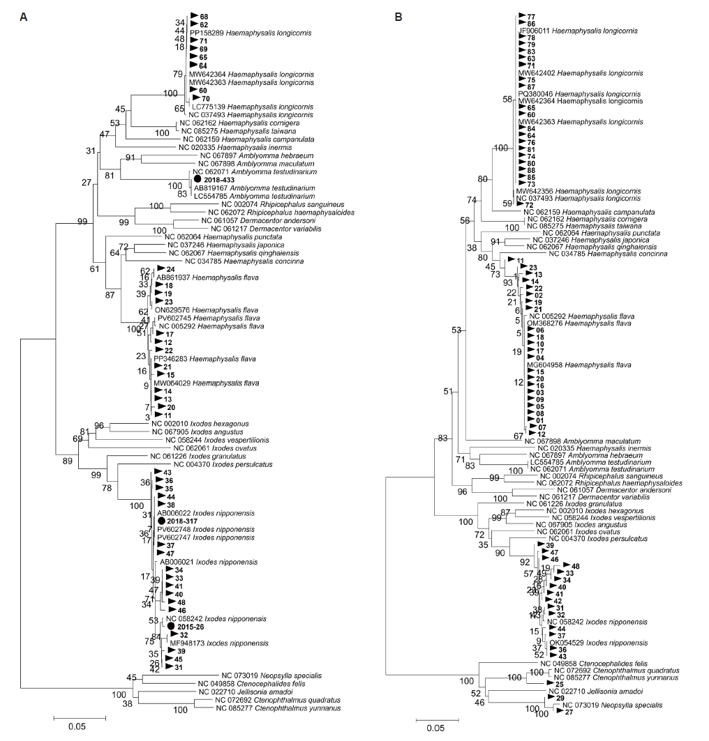
Phylogenetic analyses based on mitochondrial 16S and 12S rDNA sequences were performed to confirm species-level identification. The 16S rDNA phylogenetic tree was constructed using 41 sequences, including 38 tick specimens collected from vegetation and wild rodents and 3 damaged tick specimens removed from patients, together with reference sequences retrieved from the NCBI database. The resulting phylogenetic tree based on the 16S rDNA sequences clearly resolved
H. flava,
H. longicornis,
I. nipponensis, and
A. testudinarium into distinct and well-supported clades, with no outliers or unexpected placements (
Fig. 2A). Similarly, the 12S rDNA phylogenetic tree, constructed from 59 tick and 3 flea sequences, showed well-defined and strongly supported clades corresponding to
H. flava,
H. longicornis, and
I. nipponensis, without any anomalous branching. Additionally, sequences derived from flea specimens clustered with the reference sequences of
Ctenophthalmus sp. (
C. quadratus and
C. yunnanus Liu, Wu & Wu, 1986) and
N. specialis available in GenBank (
Fig. 2B).
Discussion
Accurate identification of tick species is essential for understanding their ecological roles, assessing the risk of tick-borne disease transmission, and developing targeted control measures [
8,
9]. In this study, we evaluated the utility of mitochondrial 16S and 12S rDNA markers in identifying hard ticks and rodent-associated ectoparasites collected from diverse sources, including vegetation, wild rodents, and patients in Gwangju, Korea.
When the specimens were morphologically intact, molecular identification using both the 16S and 12S rDNA markers yielded results consistent with those of traditional taxonomy, supporting the reliability of both approaches under optimal conditions. However, morphology-based identification is often limited by physical damage, particularly in specimens removed from human patients. In such cases, molecular analysis proved essential and 3 morphologically unidentifiable specimens were successfully classified using 16S rDNA as I. nipponensis (n=2) and A. testudinarium (n=1), demonstrating the diagnostic utility of molecular methods in clinical or degraded samples. These findings further highlight the practical value of mitochondrial markers in identifying degraded or otherwise unidentifiable tick samples when morphological features are insufficient for taxonomic discrimination.
Although both mitochondrial markers were useful for species identification, their performances varied considerably among species. The 16S rDNA marker showed a high PCR amplification rate (98.6%) but a relatively low proportion of samples yielding sequence data suitable for molecular identification (52.1%). In contrast, 12S rDNA exhibited lower amplification efficiency (86.5%) but much higher sequencing success for molecular identification (92.2%). The relatively low sequencing success of 16S rDNA, despite PCR positivity, suggests that successful amplification did not always yield sequence data of sufficient quality for reliable analysis. This may have reflected variation in amplicon quality, reduced band resolution during preparative electrophoresis for gel extraction, purification-associated loss of sequence quality, or limited sequencing readability in some samples. These differences likely reflect interspecific variations in template DNA integrity, mitochondrial genome organization, and primer-binding affinity [
12,
13].
Among the 3 tick species, I. nipponensis showed the most balanced performance across both markers, reflecting good primer compatibility and stable DNA integrity. In comparison, H. flava exhibited greater variation between the 2 loci, whereas H. longicornis showed a clear marker-dependent bias, with very low sequencing success for 16S but complete success for 12S. Together, these findings indicate that the performance of mitochondrial markers is influenced not only by marker type but also by species-specific sequence characteristics, and that marker choice may substantially affect identification outcomes. Therefore, species-specific optimization of molecular protocols is essential to enhance the accuracy and reproducibility of mitochondrial-based identification.
Previous studies have demonstrated that mitochondrial markers such as 16S and 12S rDNA are reliable tools for tick identification because of their moderate evolutionary rates, maternal inheritance, and lack of recombination [
13,
14]. The use of 12S rDNA sequences has been shown to effectively resolve phylogenetic relationships within
Rhipicephalus and
Boophilus, confirming their ability to distinguish closely related taxa [
14]. Moreover, evaluations of multiple genetic loci have confirmed that both 16S and 12S rDNA markers are suitable for accurate species discrimination in ixodid ticks [
13]. Additionally, the limitations of morphology-based identification for immature or damaged specimens have been highlighted, emphasizing the value of molecular approaches as complementary tools for species confirmation [
4].
The phylogenetic analyses conducted in this study further validated the accuracy of molecular identification. Neighbor-joining phylogenetic trees based on 16S and 12S rDNA sequences reliably distinguished H. flava, H. longicornis, I. nipponensis, and A. testudinarium, with each species forming a distinct, well-supported clade. Moreover, 12S rDNA analysis successfully identified 3 flea specimens as N. specialis (n=2) and Ctenophthalmus sp. (n=1), indicating that this marker may be applicable beyond ticks and useful for detecting other arthropod ectoparasites in rodent surveillance. However, PCR amplification failed for all lice and mite specimens, indicating that the current primers are insufficient for broad ectoparasite detection. This finding suggests that, under the current assay conditions, the markers are more suitable for targeted tick identification than for broader ectoparasite screening. Taken together, these results support the exploratory utility of 12S rDNA for selected rodent-associated ectoparasites while also underscoring the limited taxonomic breadth of the present assay conditions. This limitation underscores the need to develop degenerate primers or adopt alternative molecular targets to improve the taxonomic coverage in future surveillance studies.
The ecological context also influenced the species composition of the ticks collected. Most
H. longicornis and
H. flava specimens were collected from vegetation in adjacent urban sites, whereas
I. nipponensis was primarily associated with wild rodents, which is consistent with previous findings [
17,
18]. All
H. longicornis and
I. nipponensis specimens in this study were female, consistent with nationwide surveys reporting female dominance in field-collected ticks [
19,
20]. This female-biased pattern has also been documented in other ixodid tick populations and is thought to reflect behavioral and ecological differences between sexes. Adult females generally remain attached to hosts for extended feeding periods to complete engorgement and egg development, whereas males detach shortly after mating or exhibit limited feeding activity, resulting in their lower capture frequency during flagging or host-associated sampling [
21,
22]. Such biological differences likely contribute to the predominance of females observed in both vegetation-derived and rodent-derived samples. Importantly, this study demonstrated the presence of ticks capable of transmitting pathogens in commonly encountered environments, such as urban parks and nearby forested areas, supporting concerns regarding tick exposure risks during outdoor activities [
23-
25].
These findings also have practical implications for routine surveillance and clinical diagnostics. Mitochondrial 16S and 12S rDNA markers may complement morphological identification, particularly when tick specimens are damaged or difficult to classify, such as in field-collected or patient-derived specimens. In addition, the combined use of morphological and molecular methods may improve the accuracy of species-level data used in vector monitoring programs, thereby supporting more reliable epidemiological risk assessment and vector control planning.
Although this study focused on adult ticks, previous studies have demonstrated that the same molecular markers can be applied to larval and nymphal stages [
8,
13]. Several limitations of this study should also be considered. The number of tick specimens obtained directly from patients was small, which limits broader evaluation of the diagnostic utility of these markers in clinical specimens. Furthermore, because all specimens were collected in Gwangju, Korea, the results may not fully reflect tick diversity or marker performance in other geographic regions. The current primer sets also failed to amplify lice and mite specimens collected from wild rodents, suggesting that the current assay conditions are not yet suitable for broader ectoparasite detection without further optimization. Although the exact cause could not be determined from the current data, limited primer compatibility is a plausible explanation, while reduced DNA quality in these specimens cannot be excluded. Future studies should extend molecular evaluations to immature stages, include larger numbers of patient-derived specimens from broader geographic areas, and assess additional markers, such as COI and ITS-2, after further optimization, to improve diagnostic resolution and taxonomic coverage.
In conclusion, mitochondrial 16S and 12S rDNA markers are effective tools for species-level identification of hard ticks, particularly when morphological identification is limited. Their combined use with morphological examination may improve species confirmation in routine surveillance and clinical settings, especially for damaged or morphologically ambiguous specimens. Continued efforts to refine molecular protocols, expand reference databases, and assess broader ectoparasite taxa across wider geographic regions and specimen types are essential to enhance tick surveillance and disease risk assessment.
Notes
-
Data availability
Sequence data and other study materials are available from the corresponding author upon reasonable request.
-
Author contributions
Conceptualization: Kim DM. Data curation: Kim SG, Lee YM. Formal analysis: Kim CM, Kim SG. Funding acquisition: Kim CM. Investigation: Lee YM, Kim SG. Methodology: Kim CM, Lee YM. Project administration: Kim DM. Resources: Kim SG, Seo JW, Kim DY, Yun NR. Supervision: Seo JW, Kim DY, Yun NR. Validation: Seo JW, Kim DY, Yun NR, Kim DM. Visualization: Kim SG, Lee YM. Writing – original draft: Kim CM, Kim SG. Writing – review & editing: Kim CM, Kim DM.
-
Conflict of interest
The authors have no conflicts of interest to declare.
-
Funding
This study was supported by a research fund from Chosun University, 2021.
Fig. 1.Dorsal and ventral views of morphologically identified adult ticks (Haemaphysalis longicornis, Haemaphysalis flava, and Ixodes nipponensis), a Ctenophthalmus sp. flea collected from a wild rodent, and 3 damaged tick specimens removed from patients in this study. H. longicornis female, (A) dorsal and (B) ventral views; H. flava male, (C) dorsal and (D) ventral views; H. flava female, (E) dorsal and (F) ventral views; I. nipponensis female, (G) dorsal and (H) ventral views;(I) Ctenophthalmus sp. flea collected from a wild rodent; (J) Tick specimens removed from patients that were partially destroyed and unidentifiable by morphology: (a) severely damaged tick; (b) partially damaged tick tentatively identified as I. nipponensis; (c) partially damaged tick tentatively identified as Amblyomma testudinarium.
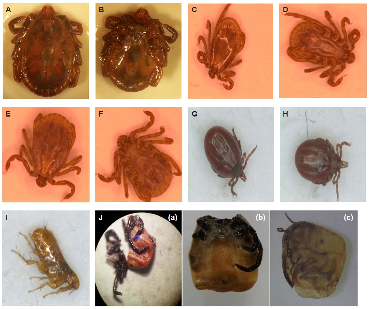
Fig. 2.Phylogenetic trees based on partial 16S and 12S ribosomal DNA (rDNA) sequences from ticks (Haemaphysalis longicornis, Haemaphysalis flava, Ixodes nipponensis, and Amblyomma testudinarium) and flea specimens (Ctenophthalmus sp. and Neopsylla specialis) collected in this study and reference sequences obtained from GenBank. Phylogenetic analyses were conducted using the neighbor-joining method based on aligned partial sequences of (A) 16S (354 bp) and (B) 12S (300 bp) rDNA. Bootstrap values (1,000 replicates) are shown at the nodes. Sequences generated in this study are marked with triangles (▶), and sequences obtained from ticks removed from human patients (n=3) are indicated by filled circles (●). GenBank accession numbers are indicated for reference sequences.
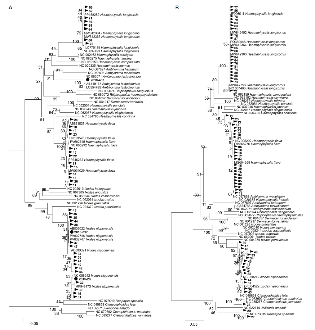
Table 1.Primer sequences used for amplification of 16S rDNA and 12S rDNA
Table 1.
|
Target gene |
Primer name |
Sequence (5′–3′) |
Expected product size (bp) |
References |
|
16S rDNA |
16s+1F |
CTG CTC AAT GAA TAT TTA AAT TGC |
450 |
[12] |
|
16S-1R |
CGG TCT AAA CTC AGA TCA TGT AGG |
|
12S rDNA |
T1B |
AAA CTA GGA TTA GAT ACC CT |
360 |
[14] |
|
T2A |
AAT GAG AGC GAC GGG CGA TGT |
Table 2.PCR amplification and sequencing results for molecular identification of tick and rodent-associated ectoparasites based on 16S and 12S rDNA
Table 2.
|
Morphological identificationa
|
PCR amplification |
Sequencing success for identification |
|
16S rDNA |
12S rDNA |
16S rDNA |
12S rDNA |
|
Haemaphysalis flava (n=24) |
24/24 (100) |
24/24 (100) |
13/24 (54.2) |
23/24 (95.8) |
|
Haemaphysalis longicornis (n=30) |
30/30 (100) |
21/30 (70.0) |
8/30 (26.7) |
21/21 (100) |
|
Ixodes nipponensis (n=20) |
19/20 (95.0) |
19/20 (95.0) |
17/19 (89.5) |
15/19 (78.9) |
|
Suspect rat flea (n=5) |
0/5 (0) |
3/5 (60.0) |
0/0 (0) |
3/3 (100) |
|
Suspect rat mite (n=5) |
0/5 (0) |
0/5 (0) |
0/0 (0) |
0/0 (0) |
|
Suspect rat louse (n=5) |
0/5 (0) |
0/5 (0) |
0/0 (0) |
0/0 (0) |
References
- 1. World Health Organization. Vector-borne diseases [Internet]. 2024 [cited 2026 Feb 1]. Available from: https://www.who.int/news-room/fact-sheets/detail/vector-borne-diseases
- 2. Song BG, Lee WG, Ju YR. Geographical distribution of Ixodid ticks in the Republic of Korea, 2015. Public Health Wkly Rep 2017;10:107-113.
- 3. Gai ZT, Zhang Y, Liang MF, et al. Clinical progress and risk factors for death in severe fever with thrombocytopenia syndrome patients. J Infect Dis 2012;206:1095-102. https://doi.org/10.1093/infdis/jis472
- 4. Walker AR, Bouattour A, Camicas JL, et al. Ticks of domestic animals in Africa: a guide to identification of species. University of Edinburgh; 2003.
- 5. Yu XJ, Liang MF, Zhang SY, et al. Fever with thrombocytopenia associated with a novel bunyavirus in China. N Engl J Med 2011;364:1523-32. https://doi.org/10.1056/NEJMoa1010095
- 6. Park SW, Song BG, Shin EH, et al. Prevalence of severe fever with thrombocytopenia syndrome virus in Haemaphysalis longicornis ticks in South Korea. Ticks Tick Borne Dis 2014;5:975-7. https://doi.org/10.1016/j.ttbdis.2014.07.020
- 7. Luo LM, Zhao L, Wen HL, et al. Haemaphysalis longicornis ticks as reservoir and vector of severe fever with thrombocytopenia syndrome virus in China. Emerg Infect Dis 2015;21:1770-6. https://doi.org/10.3201/eid2110.150126
- 8. Eisen RJ, Paddock CD. Tick and tickborne pathogen surveillance as a public health tool in the United States. J Med Entomol 2021;58:1490-502. https://doi.org/10.1093/jme/tjaa087
- 9. Makwarela TG, Seoraj-Pillai N, Nangammbi TC. Tick control strategies: critical insights into chemical, biological, physical, and integrated approaches for effective hard tick management. Vet Sci 2025;12:114. https://doi.org/10.3390/vetsci12020114
- 10. Intirach J, Lv X, Han Q, Lv ZY, Chen T. Morphological and molecular identification of hard ticks in Hainan Island, China. Genes (Basel) 2023;14:1592. https://doi.org/10.3390/genes14081592
- 11. Makwarela TG, Nyangiwe N, Masebe TM, et al. Morphological and molecular characterization of tick species infesting cattle in South Africa. Vet Sci 2024;11:638. https://doi.org/10.3390/vetsci11120638
- 12. Tian Z, Liu G, Xie J, et al. Discrimination between Haemaphysalis longicornis and H. qinghaiensis based on the partial 16S rDNA and the second internal transcribed spacer (ITS-2). Exp Appl Acarol 2011;54:165-72. https://doi.org/10.1007/s10493-010-9423-3
- 13. Lv J, Wu S, Zhang Y, et al. Assessment of four DNA fragments (COI, 16S rDNA, ITS2, 12S rDNA) for species identification of the Ixodida (Acari: Ixodida). Parasit Vectors 2014;7:93. https://doi.org/10.1186/1756-3305-7-93
- 14. Beati L, Keirans JE. Analysis of the systematic relationships among ticks of the genera Rhipicephalus and Boophilus (Acari: Ixodidae) based on mitochondrial 12S ribosomal DNA gene sequences and morphological characters. J Parasitol 2001;87:32-48. https://doi.org/10.1645/0022-3395(2001)087[0032:AOTSRA]2.0.CO;2
- 15. Jalal S, Jha B, Kim CM, et al. Molecular detection of viruses causing hemorrhagic fevers in rodents in the south-west of Korea. J Neurovirol 2019;25:239-47. https://doi.org/10.1007/s13365-018-0708-3
- 16. Yamaguti N, Tipton VJ, Keegan HL, Toshioka S. Ticks of Japan, Korea, and the Ryukyu Islands. Brigh Young Univ Sci Bull Biol Ser 1971;15:1.
- 17. Kim CM, Yi YH, Yu DH, et al. Tick-borne rickettsial pathogens in ticks and small mammals in Korea. Appl Environ Microbiol 2006;72:5766-76. https://doi.org/10.1128/AEM.00431-06
- 18. Kim HC, Han SH, Chong ST, et al. Ticks collected from selected mammalian hosts surveyed in the Republic of Korea during 2008-2009. Korean J Parasitol 2011;49:331-5. https://doi.org/10.3347/kjp.2011.49.3.331
- 19. Kim-Jeon MD, Jegal S, Jun H, et al. Four year surveillance of the vector hard ticks for SFTS, Ganghwa-do, Republic of Korea. Korean J Parasitol 2019;57:691-8. https://doi.org/10.3347/kjp.2019.57.6.691
- 20. Kang SW, Doan HT, Choe SE, et al. Molecular investigation of tick-borne pathogens in ticks from grazing cattle in Korea. Parasitol Int 2013;62:276-82. https://doi.org/10.1016/j.parint.2013.02.002
- 21. Finney SK, Elston DA. Natural attachment duration of adult female ticks Ixodes uriae (Acari: Ixodidae) on free-living adult black-legged kittiwakes Rissa tridactyla. Exp appl acarol 1999;23:765-9. https://doi.org/10.1023/A:1006249309647
- 22. Richards SL, Langley R, Apperson CS, Watson E. Do tick attachment times vary between different tick-pathogen systems? Environments 2017;4:37. https://doi.org/10.3390/environments4020037
- 23. Niu G, Li J, Liang M, et al. Severe fever with thrombocytopenia syndrome virus among domesticated animals, China. Emerg Infect Dis 2013;19:756-63. https://doi.org/10.3201/eid1905.120245
- 24. Liang S, Bao C, Zhou M, et al. Seroprevalence and risk factors for severe fever with thrombocytopenia syndrome virus infection in Jiangsu Province, China, 2011. Am J Trop Med Hyg 2014;90:256-9. https://doi.org/10.4269/ajtmh.13-0423
- 25. Kato CY, Chung IH, Robinson LK, et al. Assessment of real-time PCR assay for detection of Rickettsia spp. and Rickettsia rickettsii in banked clinical samples. J Clin Microbiol 2013;51:314-7. https://doi.org/10.1128/JCM.01723-12


 , Seon Gyeong Kim2,†, You Mi Lee3