Abstract
Trypanosoma brucei, the protozoan parasite responsible for African trypanosomiasis, undergoes a divergent cell cycle characterized by closed mitosis, inheritance of single-copy organelles such as the kinetoplast, basal body, and flagellum, and anterior-to-posterior longitudinal cytokinesis. Cdc2-related kinases (CRKs) and cyclin-like proteins have emerged as key regulators of these processes; however, most CRK–cyclin modules remain incompletely defined in terms of their in vivo composition, substrate specificity, and physiological function. In this review, we summarize current evidence for the roles of T. brucei CRKs in parasite cell-cycle control, focusing on CRK1-associated PHO80-like cyclins in G1/S progression, the CRK2–CYC13 complex in S-phase DNA replication, CRK3-associated complexes in mitosis, and additional modules including CRK9–CYC12 in spliced-leader trans-splicing and CRK12–CYC9 with endocytic and cytokinesis-related phenotypes. By comparing evidence across these modules, we highlight the gap between phenotypic assignment and mechanistic validation, and emphasize the need to define functional CRK–cyclin–substrate axes in vivo. Resolving these gaps will provide important insights into parasite-specific cell-cycle control and support the evaluation of CRKs as parasite-selective therapeutic targets.
-
Key words: Trypanosoma brucei, Cdc2-related kinase, cyclins, cell cycle, DNA replication, mitosis
Introduction
Trypanosoma brucei is an early-branching unicellular eukaryotic parasite whose subspecies cause human African trypanosomiasis (HAT), or sleeping sickness, in sub-Saharan Africa [
1]. More broadly, tsetse fly (
Glossina spp.)-transmitted African trypanosomes, including
T. brucei, impose a dual burden on human health and livestock productivity, making trypanosomiasis a representative One Health issue in the region [
1,
2]. Although sustained control efforts have markedly reduced reported HAT cases, the disease remains classified by the World Health Organization as a neglected tropical disease, and animal African trypanosomiasis, or nagana, continues to constrain livestock production in affected areas [
1,
2].
Chemotherapy against HAT has improved, but important limitations remain. The oral nitroimidazole fexinidazole has simplified treatment for many HAT cases, and recent World Health Organization guidelines have expanded its use [
3-
5]; nevertheless, therapeutic decisions still depend on the causative trypanosome, disease stage, patient eligibility, and access to available regimens [
5]. While target-based approaches have advanced in parallel, phenotypic screening has been widely used in neglected tropical disease drug discovery to identify compounds that inhibit parasite growth [
6]. However, without target deconvolution and mechanistic characterization, hit optimization, selectivity assessment, and rational drug development remain challenging [
7]. The absence of an effective prophylactic vaccine further reinforces the need to identify parasite-selective molecular targets for mechanism-based drug development [
1,
8].
A critical basis for such parasite selectivity lies in the divergent cell biology of
T. brucei. Although the parasite retains core eukaryotic cell-cycle principles, proliferation requires the ordered duplication and segregation of specialized structures, including the flagellum–basal body complex, the flagellum attachment zone (FAZ), and the kinetoplast. These events must be coordinated with nuclear DNA replication, mitosis, and longitudinal cytokinesis [
9-
12]. Defining how parasite-specific cell-cycle regulators coordinate nuclear and extranuclear inheritance events in time and space should provide important insight into
T. brucei cell-cycle control and help evaluate these regulators as selective therapeutic targets.
In most well-studied eukaryotes, cell-cycle progression is governed by cyclin-dependent kinases (CDKs) and their cyclin partners.
T. brucei possesses a distinctive repertoire of Cdc2-related kinases (CRKs) and cyclin-like proteins [
13,
14], including PHO80-like cyclins implicated in G1/S regulation [
15]. Its genome encodes eleven CRKs, designated CRK1–CRK4 and CRK6–CRK12 [
13,
14], although these family members differ substantially in the extent to which their cyclin partners, substrates, and physiological functions have been defined. Functional studies have linked several CRKs to distinct cellular processes, including CRK1 in G1/S progression, CRK2 in S-phase DNA replication, CRK3 in G2/M regulation, CRK9 in spliced-leader trans-splicing, and CRK12 in endocytosis- and cytokinesis-associated phenotypes [
16-
20]. However, for many CRK–cyclin modules, their precise composition, activation state, and physiological substrates remain incompletely defined, creating a critical gap between phenotypic observation and mechanistic understanding. A well-resolved CRK–cyclin–substrate map would help clarify how the parasite couples DNA replication, organelle segregation, cytokinesis, and RNA-processing-associated regulation, while also offering a rational framework for evaluating CRKs as parasite-selective kinase targets.
In this review, we summarize and critically evaluate current evidence for
T. brucei CRKs and their cyclin partners as regulators of parasite cell-cycle progression, focusing on CRK1-associated PHO80-like cyclins, CRK2–CYC13, CRK3-associated complexes, CRK9–CYC12, and CRK12–CYC9. Rather than treating these modules as equally resolved, we compare the strength of evidence supporting their cyclin associations, proposed substrates, and physiological functions. Key features of these modules are summarized in
Table 1 [
15-
27].
The Unique Cell-cycle Architecture of T. brucei
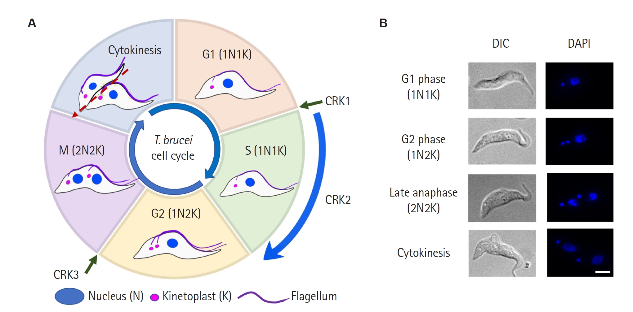
At a broad level,
T. brucei follows the canonical eukaryotic sequence of G1, S phase, G2, mitosis, and cytokinesis [
11,
12,
14]. However, the cellular execution of this sequence differs substantially from the animal-cell paradigm and is shaped by the parasite's polarized morphology and single-copy organelles. First,
T. brucei undergoes closed mitosis: the nuclear envelope remains intact, and the mitotic spindle is assembled within the nucleus rather than after nuclear envelope breakdown [
11,
12]. Second, cell-cycle progression requires not only nuclear DNA replication and chromosome segregation, but also the ordered duplication and partitioning of the basal body, flagellum, FAZ, and the kinetoplast—the mitochondrial DNA network [
9,
11,
12,
28]. These events are commonly monitored by classifying cells according to their nuclear and kinetoplast content: cells with 1 nucleus and 1 kinetoplast (1N1K), cells with 1 nucleus and 2 kinetoplasts (1N2K), and cells with 2 nuclei and 2 kinetoplasts (2N2K). In this scheme, N denotes the nucleus and K denotes the kinetoplast [
9,
11]. Importantly, kinetoplast replication begins slightly before nuclear S phase, and basal body duplication and separation are tightly coupled to kinetoplast segregation [
9,
10,
12].
Cytokinesis also differs markedly from that of metazoan cells.
T. brucei lacks a conventional actomyosin contractile ring, and cleavage furrow positioning is linked to the elongating new FAZ and flagellum-associated cytoskeletal structures [
11,
12,
28,
29]. The furrow ingresses longitudinally from the anterior toward the posterior pole rather than by equatorial constriction [
11,
12,
28,
29]. These features provide an essential framework for interpreting CRK loss-of-function phenotypes, which may involve altered DNA content, abnormal N/K configurations, defective kinetoplast segregation, flagellum/FAZ morphogenesis defects, or cytokinesis failure (
Fig. 1).
CRK1 and PHO80-like Cyclins in the G1/S Transition
In eukaryotic cells, the G1/S transition represents a critical control point at which cells commit to DNA replication and subsequent cell-cycle progression. During G1 phase, cells integrate growth status, nutrient availability, mitogenic cues, and DNA damage signals before passing start or the restriction point and entering S phase. This transition is primarily governed by CDK–cyclin complexes, whose stage-specific activation promotes DNA replication initiation, transcriptional reprogramming, and checkpoint control. Accordingly, accurate regulation of CDK activity at the G1/S boundary is essential for proper cell proliferation and genome stability [
30,
31]. In
T. brucei, this regulatory problem is further complicated by the parasite-specific cellular architecture introduced above, which couples nuclear DNA replication to posterior cytoskeletal morphogenesis [
32,
33].
Among the CRKs in
T. brucei, CRK1 has emerged as a key regulator of G1/S progression. Early RNAi and pairwise knockdown studies linked CRK1 depletion to G1/S defects and CRK3 depletion to G2/M defects [
16,
22]. Notably, simultaneous depletion of CRK1 and CRK2 caused a stronger G1 arrest than either knockdown alone, accompanied by inhibition of nuclear DNA synthesis and abnormal posterior-end elongation or branching [
22,
33]. These phenotypes highlight the coupling between G1/S commitment and posterior cytoskeletal morphogenesis. The later identification of the CRK2–CYC13 complex as an S-phase CDK-like module involved in DNA replication initiation further refined this interpretation [
17], suggesting that the phenotype observed after simultaneous depletion of CRK1 and CRK2 may reflect convergent defects in G1/S commitment and replication initiation. In parallel, several PHO80-like cyclins, including CYC2, CYC4, CYC5, and CYC7, have been proposed as CRK1-associated regulatory partners [
15,
27], based on interaction assays and RNAi phenotypes linking their depletion to defects in cell-cycle progression, morphology, and posterior cytoskeletal organization. Thus, current evidence supports a CRK1–PHO80-like cyclin module that promotes G1/S progression and cytoskeletal morphogenesis, but the precise in vivo composition, activation timing, and physiological substrates of this module remain unresolved [
14,
15,
23,
24].
Downstream of CRK1, several candidate phosphorylation targets, including Sec31, eIF4E4, and PABP1, have been reported [
23,
24], although their directness and the responsible CRK1–cyclin complexes in vivo remain incompletely defined. CRK1-mediated phosphorylation of the coat protein complex II subunit Sec31 links CRK1 activity to anterograde protein transport [
23], whereas phosphorylation of eIF4E4 and PABP1 implicates CRK1 in cap-dependent translation initiation [
24]. These substrate classes suggest that CRK1 may coordinate G1/S progression with broader biosynthetic programs, rather than acting solely as a direct trigger of DNA replication. However, the evidence supporting these CRK1-linked substrates does not yet reach the mechanistic depth established for the CRK2–CYC13–Mcm3 axis, in which phospho-site mapping and phospho-mutant rescue together support the physiological relevance of phosphorylation [
17]. Thus, although CRK1 has been linked to both secretory and translational pathways, the assignment of these phosphorylation events to specific CRK1–cyclin complexes and their precise contribution to G1/S progression remain unresolved. A key question is whether these substrates directly mediate G1/S commitment or instead support the biosynthetic capacity required for cell-cycle entry. Additional, as-yet-unidentified CRK1 substrates may also connect CRK1 activity more directly to the G1/S transition. Defining the relevant CRK1–PHO80-like cyclin complex, its activation timing, and its substrate hierarchy will therefore be necessary to distinguish direct G1/S regulatory functions from indirect effects on secretion, translation, or cellular growth.
CRK2–CYC13 Mediated Regulation of DNA Replication
In eukaryotic cells, S phase requires the once-per-cell-cycle duplication of nuclear DNA before chromosome segregation. Replication initiation is controlled by conserved licensing and firing mechanisms, in which pre-replicative complexes assembled in G1 are converted into active Cdc45–Mcm2–7–GINS (CMG) helicases at the onset of S phase [
30,
31]. In
T. brucei, however, this regulatory system is highly divergent [
34,
35]: the parasite possesses a conserved CMG complex but lacks close homologues of several canonical replication regulators, including Cdc6, Cdt1, Sld2, Sld3, and Dbf4-dependent kinase (DDK)/Cdk7 [
34,
36,
37]. The absence of Sld2 and Sld3 is noteworthy, because S-phase CDK–dependent phosphorylation of these factors provides the canonical route for GINS recruitment and CMG activation in other eukaryotes [
30,
31]. This divergence raises the question of how
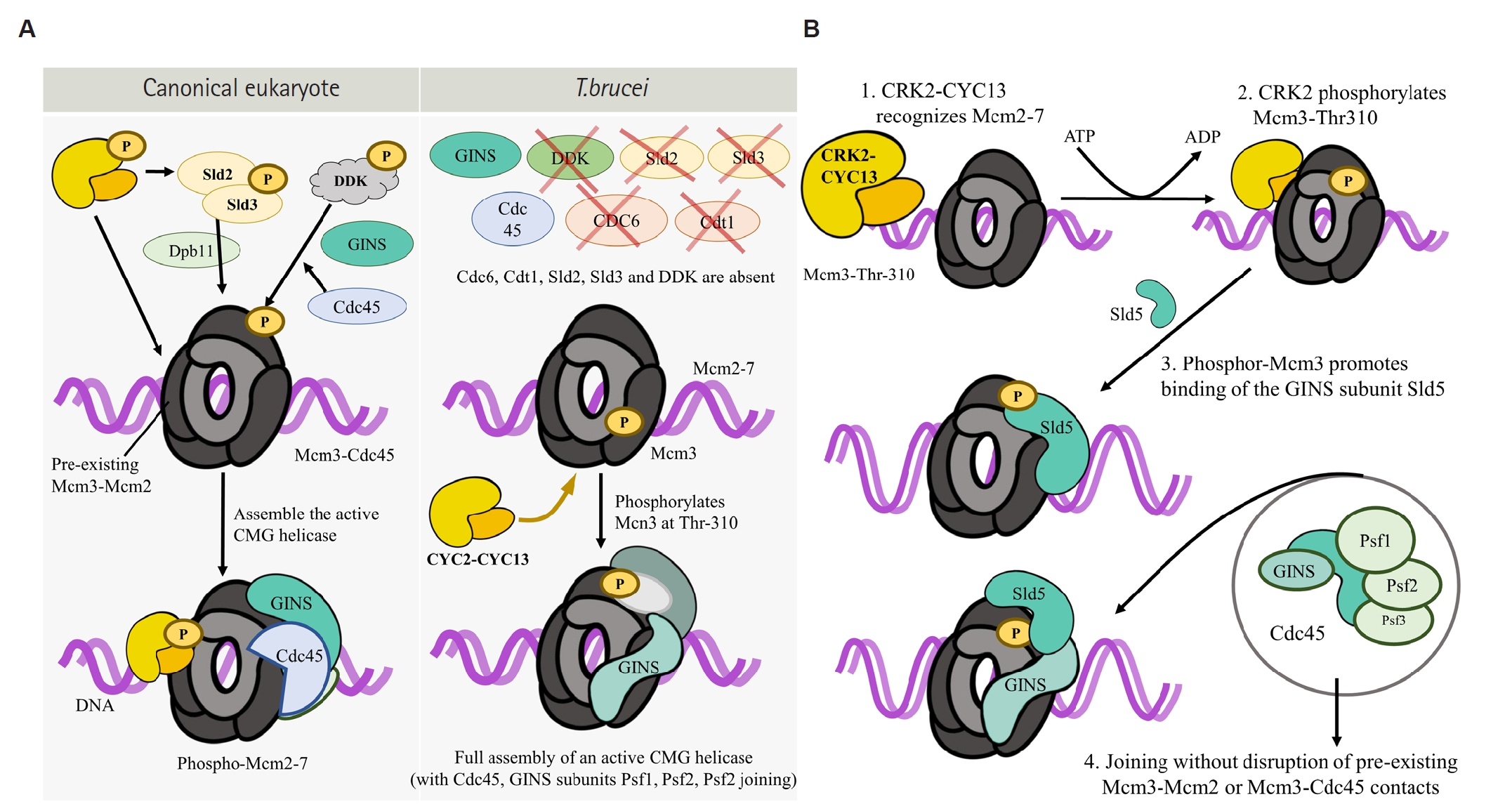
T. brucei couples Mcm2–7 loading to GINS recruitment and CMG activation in the absence of the conventional Sld2/Sld3-dependent pathway (
Fig. 2A).
Earlier RNAi approaches achieved only partial depletion of CRK2, leaving its cell-cycle function less clearly resolved [
16,
22]. Subsequent work, however, using an efficient tetracycline-inducible RNAi strategy achieved more than 90% depletion of CRK2 and CYC13 at the protein level, thereby demonstrating that both proteins are essential for cell proliferation [
17]. Although yeast two-hybrid assays suggested that CYC13 can interact with multiple CRKs, reciprocal co-immunoprecipitation demonstrated that CYC13 forms an in vivo complex specifically with CRK2 [
17]. Functionally, depletion of either CRK2 or CYC13 caused severe growth defects and impaired DNA replication, as shown by S-phase accumulation and reduced EdU incorporation in 1N1K cells [
17]. These findings established CRK2–CYC13 as a major S-phase CDK-like module in
T. brucei. However, CYC13 depletion produced a broader phenotype than CRK2 depletion, including cells with DNA content greater than 4N and anucleate cells, suggesting that CYC13 may have additional CRK2-independent functions [
17].
The CRK2–CYC13 study moved beyond phenotypic assignment by identifying Mcm3, a subunit of the Mcm2–7 helicase, as a mechanistic substrate of CRK2 (
Fig. 2A) [
17]. CRK2 phosphorylates Mcm3 in a CYC13-enhanced manner, with Thr-310 emerging as a functionally important phosphorylation site: phospho-deficient Mcm3-T310A failed to restore cell proliferation and DNA replication, whereas phosphomimetic Mcm3-T310D partially rescued these defects [
17]. Mechanistically, CRK2-dependent Mcm3 phosphorylation promotes Mcm3 association with Sld5, a GINS subunit, without disrupting Mcm3 interaction with Mcm2 or Cdc45 [
17]. These findings suggest that CRK2–CYC13 supports DNA replication initiation by facilitating GINS association with Mcm3 and thereby promoting assembly or stabilization of the CMG helicase (
Fig. 2B) [
17].
Together, these results make CRK2–CYC13–Mcm3 the most mechanistically resolved CRK–cyclin–substrate module currently defined in T. brucei, because cyclin association, substrate identification, phosphorylation-site mapping, and functional rescue analyses have been connected within a single pathway. More broadly, this pathway suggests that CRK2-mediated Mcm3 phosphorylation may provide an alternative route for promoting GINS association with Mcm2–7 in the absence of canonical Sld2/Sld3-dependent recruitment. Thus, CRK2–CYC13 may represent a parasite-specific solution to a conserved replication problem rather than a simple one-to-one replacement of canonical S-phase CDK pathways.
Despite this advance, several questions remain unresolved. Although Mcm3 is a validated CRK2 substrate [
17], it remains unknown whether CRK2–CYC13 phosphorylates additional substrates involved in replication initiation, fork progression, or replication-stress responses. Moreover, the broader phenotype caused by CYC13 depletion, including cells with post-G2/M DNA content and anucleate cells, raises the possibility that CYC13 may act with other CRKs or participate in CRK2-independent cell-cycle processes. Finally, because
T. brucei lacks canonical DDK/Cdk7 homologues [
17,
34], it remains unclear whether CRK2–CYC13 substitutes only for an S-phase CDK-like function or also compensates for regulatory steps that are divided between DDK and S-phase CDKs in other eukaryotes. In addition, although the CRK2–CYC13–Mcm3 pathway provides a plausible mechanism for promoting GINS association with Mcm2–7, how Cdc45 is recruited to the helicase complex in this divergent system remains unresolved. Thus, CRK2–CYC13 provides a benchmark for evaluating other
T. brucei CRK–cyclin modules, while also highlighting the divergent logic of DNA replication control in this parasite.
CRK3-associated Cyclin Complexes in Mitotic Progression
CRK3 has been most closely associated with G2/M progression and mitotic control in
T. brucei [
16,
21,
22]. Early RNAi studies showed that CRK3 depletion produces defects consistent with impaired G2/M transition, clearly distinguishing its functional profile from that of CRK1, which is primarily linked to G1/S progression [
16,
22]. Subsequent studies identified several CRK3-associated cyclin interactions, including CRK3–CYC2 and CRK3–CYC6 [
21,
26]. Among these, CYC6 provides the strongest support for a functional mitotic CRK3 complex: CYC6 physically interacts with CRK3, supports CRK3-associated kinase activity, and is required for mitotic progression, particularly in the procyclic form [
21].
Biochemical evidence for CRK3-associated kinase activity has largely relied on in vitro histone H1 kinase assays, together with interaction assays such as yeast two-hybrid analysis and co-immunoprecipitation [
21,
26]. These studies established that CRK3 can associate with cyclin partners and form catalytically active kinase complexes. However, because histone H1 serves as an artificial assay substrate, this activity should be interpreted as evidence of catalytic potential rather than definitive proof of endogenous substrate phosphorylation in vivo. Thus, the physiological substrates through which CRK3 controls mitotic entry, chromosome segregation, or subsequent cell-cycle events remain unidentified.
Compared with the recently elucidated CRK2–CYC13–Mcm3 replication axis (
Fig. 2), CRK3-associated modules therefore remain less mechanistically resolved. Although CRK3 is strongly supported as a regulator of G2/M progression and mitotic control, the precise composition, temporal activation, and physiological substrates of CRK3-associated complexes still need to be defined. Resolving this substrate gap will be essential for understanding how CRK3-associated complexes coordinate mitotic entry, chromosome segregation, and the transition from mitosis to cytokinesis in
T. brucei. Future work should prioritize the identification of endogenous CRK3 substrates that control mitotic progression and the transition to cytokinesis, using approaches such as stage-resolved phosphoproteomics, endogenous complex purification, chemical-genetic kinase inhibition, and phospho-mutant rescue. Such studies would also provide a clearer basis for evaluating CRK3 as a parasite-selective mitotic kinase target. The translational relevance of this gap is underscored by inhibitor-screening studies against
Leishmania CRK3, which supports broader drug-target interest in trypanosomatid mitotic kinases while also emphasizing the need for parasite-specific validation in
T. brucei [
38,
39].
Additional CRK–cyclin Modules
Beyond the major CRK modules implicated in G1/S progression, S-phase DNA replication, and G2/M transition, several additional CRKs have been investigated in
T. brucei, although their links to canonical cell-cycle transitions remain less direct or less mechanistically defined. CRK4 and CRK6 were examined in early RNAi and pairwise knockdown studies together with CRK1, CRK2, and CRK3; however, these analyses primarily reinforced the assignment of CRK1 to G1/S progression and CRK3 to G2/M control rather than defining specific roles for CRK4 or CRK6 [
16,
22]. For this reason, CRK4 and CRK6 are not included as separate modules in the summary table (
Table 1) [
15-
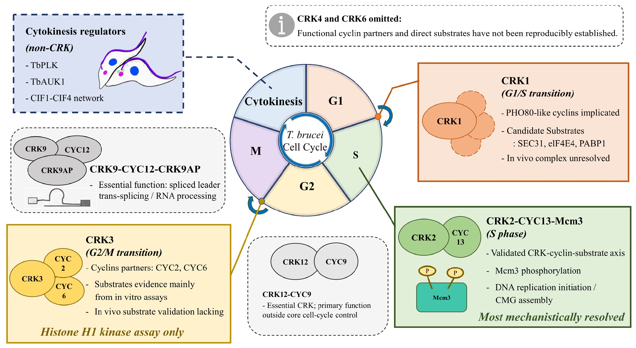
27] and integrative map (
Fig. 3), as their functional cyclin partners, activation timing, and physiological substrates remain unresolved [
14]. In contrast, 2 additional CRK–cyclin complexes, CRK9–CYC12 and CRK12–CYC9, have been characterized in
T. brucei, although their links to canonical cell-cycle transitions remain indirect [
18-
20].
CRK9 provides an instructive example of an essential CRK whose primary function appears to lie outside canonical cell-cycle transition control. Although CRK9 depletion was initially associated with defects in parasite proliferation and cell-cycle progression [
25], subsequent studies demonstrated that CRK9 is essential for spliced-leader trans-splicing, a kinetoplastid-specific mode of pre-mRNA maturation [
18,
19]. CRK9 functions in a complex with the L-type cyclin CYC12 and the kinetoplastid-specific CRK9-associated protein CRK9AP, and depletion of CYC12 or CRK9AP phenocopies CRK9 depletion [
18]. Chemical-genetic inhibition of analog-sensitive CRK9 further showed that CRK9 kinase activity is required for ongoing pre-mRNA splicing [
19]. Thus, CRK9 is best viewed as an essential RNA-processing-coupled CRK module whose effects on proliferation likely arise from defective mRNA maturation rather than direct control of a discrete cell-cycle transition.
CRK12 represents another essential but incompletely resolved CRK module. CRK12 forms a complex with CYC9, and this CRK12–CYC9 complex is required for parasite proliferation [
20]. Functional characterization linked CRK12 depletion primarily to impaired endocytosis, whereas CYC9 depletion was associated with cytokinesis-related defects. This phenotypic divergence suggests that CRK12–CYC9 may influence cell-cycle progression indirectly through endocytic or trafficking-related processes, although its direct physiological substrates and precise connection to cytokinesis remain unclear. It also cautions against interpreting CRK12–CYC9 simply as a canonical cytokinesis kinase module without further substrate-level validation.
Together, these additional CRK modules indicate that cell-cycle progression in
T. brucei is intertwined not only with classical transition control but also with RNA processing, endocytic trafficking, and parasite-specific cellular organization. They also underscore the need to distinguish CRKs that directly regulate cell-cycle transitions from CRKs whose depletion secondarily produces cell-cycle defects through disruption of essential non-cell-cycle processes. This distinction is particularly important when interpreting cytokinesis-related phenotypes, because direct CRK–cyclin involvement in
T. brucei cytokinesis itself remains poorly defined. Although
T. brucei Polo-like kinase (TbPLK),
T. brucei Aurora kinase (TbAUK1), and the CIF1-containing cytokinesis initiation network have emerged as central regulators of cleavage-furrow initiation and progression [
40-
42], whether CRK–cyclin modules directly control these pathways has not been systematically established. At present, cytokinesis-related phenotypes observed after depletion of some CRKs or cyclins are difficult to distinguish from secondary consequences of earlier defects in DNA replication, mitosis, trafficking, or cytoskeletal morphogenesis. Therefore, future studies should test whether CRKs directly phosphorylate components of the CIF network, FAZ-associated proteins, or furrow-positioning regulators, rather than inferring cytokinesis regulation solely from depletion phenotypes.
Mechanistic Gaps and Future Directions
Across
T. brucei CRK biology, a central unresolved problem is how to distinguish direct CRK–cyclin substrates from secondary downstream consequences of cell-cycle disruption. RNAi phenotypes have been essential for assigning CRKs to broad cell-cycle processes, but depletion of essential kinases can rapidly produce pleiotropic defects in DNA content, organelle segregation, morphology, and cytokinesis [
15,
16,
20,
22]. Similarly, interaction assays and in vitro kinase activity demonstrate biochemical potential, but they do not by themselves establish that a CRK–cyclin pair functions as a stage-specific kinase complex on a physiological substrate inside the parasite [
15,
20,
21]. This distinction is evident in the better-studied CRK modules.
For CRK1, depletion phenotypes and candidate phosphorylation targets such as Sec31, eIF4E4, and PABP1 support a role in G1/S-associated cellular remodeling, but the responsible in vivo CRK1–cyclin complex and the direct contribution of these phosphorylation events to G1/S commitment remain unresolved [
15,
23,
24]. For CRK3, genetic and biochemical evidence supports a role in G2/M progression, yet the physiological substrates that mediate mitotic control remain unidentified [
16,
21,
22]. In contrast, the CRK2–CYC13–Mcm3 pathway currently represents the strongest mechanistic example because cyclin association, substrate identification, phosphosite mapping, and phospho-mutant rescue have been integrated into a single functional model [
17]. Thus, most CRK modules remain positioned between phenotypic assignment and full mechanistic validation.
To move beyond phenotypic assignment and establish direct CRK–cyclin–substrate relationships, future studies should combine 3 complementary approaches. First, functional CRK–cyclin complexes should be defined under endogenous cellular conditions using endogenous tagging, reciprocal co-immunoprecipitation, and, where possible, stage-resolved interaction analyses to determine when and where each complex is assembled. Second, direct substrates should be identified using quantitative phosphoproteomics, proximity-based approaches, and chemical-genetic strategies such as analog-sensitive kinase systems; the successful use of analog-sensitive CRK9 to acutely block pre-mRNA splicing illustrates the feasibility of this approach for kinetoplastid CRKs [
19]. Third, candidate phosphorylation events should be tested by phospho-deficient and phosphomimetic rescue experiments to determine whether they are required for specific cell-cycle steps rather than merely correlating with CRK depletion phenotypes, as exemplified by the Mcm3-T310 analysis in the CRK2–CYC13 module [
17].
Beyond mechanistic validation, comparative analysis across kinetoplastid species—such as Leishmania and T. cruzi—and chemical-biology screens against
T. brucei kinases could help establish whether individual CRK–cyclin modules can be exploited as parasite-selective therapeutic targets [
38,
39]. Such cross-species and chemical-genetic insights will be particularly valuable for distinguishing essential CRK functions that are shared across trypanosomatids from those that are uniquely required in
T. brucei.
Conclusion
T. brucei CRKs constitute a central regulatory layer of the parasite cell cycle and provide a framework for understanding how the parasite coordinates DNA replication, mitosis, cytokinesis, and specialized organelle inheritance. Current evidence supports distinct CRK modules acting at major cell-cycle stages, including CRK1-associated pathways in G1/S progression, the CRK2–CYC13–Mcm3 axis in DNA replication initiation, and CRK3-associated complexes in mitotic control, and CRK9- or CRK12-associated modules linked to RNA processing, endocytosis, and cytokinesis-associated phenotypes. Among these, the CRK2–CYC13–Mcm3 axis represents a particularly well-resolved example of a defined CRK–cyclin–substrate pathway, whereas many other CRK modules remain incompletely resolved in terms of their in vivo cyclin composition, activation timing, and physiological substrates (
Fig. 3). Defining the composition, activation timing, and physiological substrates of these modules in vivo will be essential for understanding parasite-specific cell-cycle control and for assessing whether CRKs can be exploited as selective therapeutic targets in African trypanosomiasis.
Notes
-
Author contributions
Conceptualization: Lee D, Lee KJ. Data curation: Lee EH, Kim H, Han JH, Lee D, Lee KJ. Funding acquisition: Lee KJ. Investigation: Lee EH. Methodology: Lee EH, Kim H, Han JH, Lee D, Lee KJ. Supervision: Lee D, Lee KJ. Visualization: Lee EH. Writing – original draft: Lee EH. Writing – review & editing: Lee D, Lee KJ.
-
Conflict of interest
Jin-Hee Han and Kyu Joon Lee serve as editors of Parasites, Hosts and Diseases but had no involvement in the decision to publish this article. No other potential conflicts of interest relevant to this study were reported.
-
Funding
This research was supported by the National Research Foundation of Korea (NRF; grant No. RS-2025-00518392) and Soonchunhyang University Research Fund.
-
Acknowledgments
The authors acknowledge the use of an artificial intelligence tool solely for language editing and improvement of manuscript readability.
Fig. 1.Overview of Trypanosoma brucei cell-cycle progression. (A) Circular map of the T. brucei cell cycle, showing G1, S, G2, mitosis, and cytokinesis with representative nucleus/kinetoplast states and the major Cdc2-related kinase (CRK) regulatory windows discussed in this review: CRK1 at the G1/S transition, CRK2 across S phase, CRK3 at the G2/M transition. (B) Representative differential interference contrast (DIC) and 4′,6-diamidino-2-phenylindole (DAPI) images showing nuclear and kinetoplast configurations during cell-cycle progression. Scale bar=5 μm.
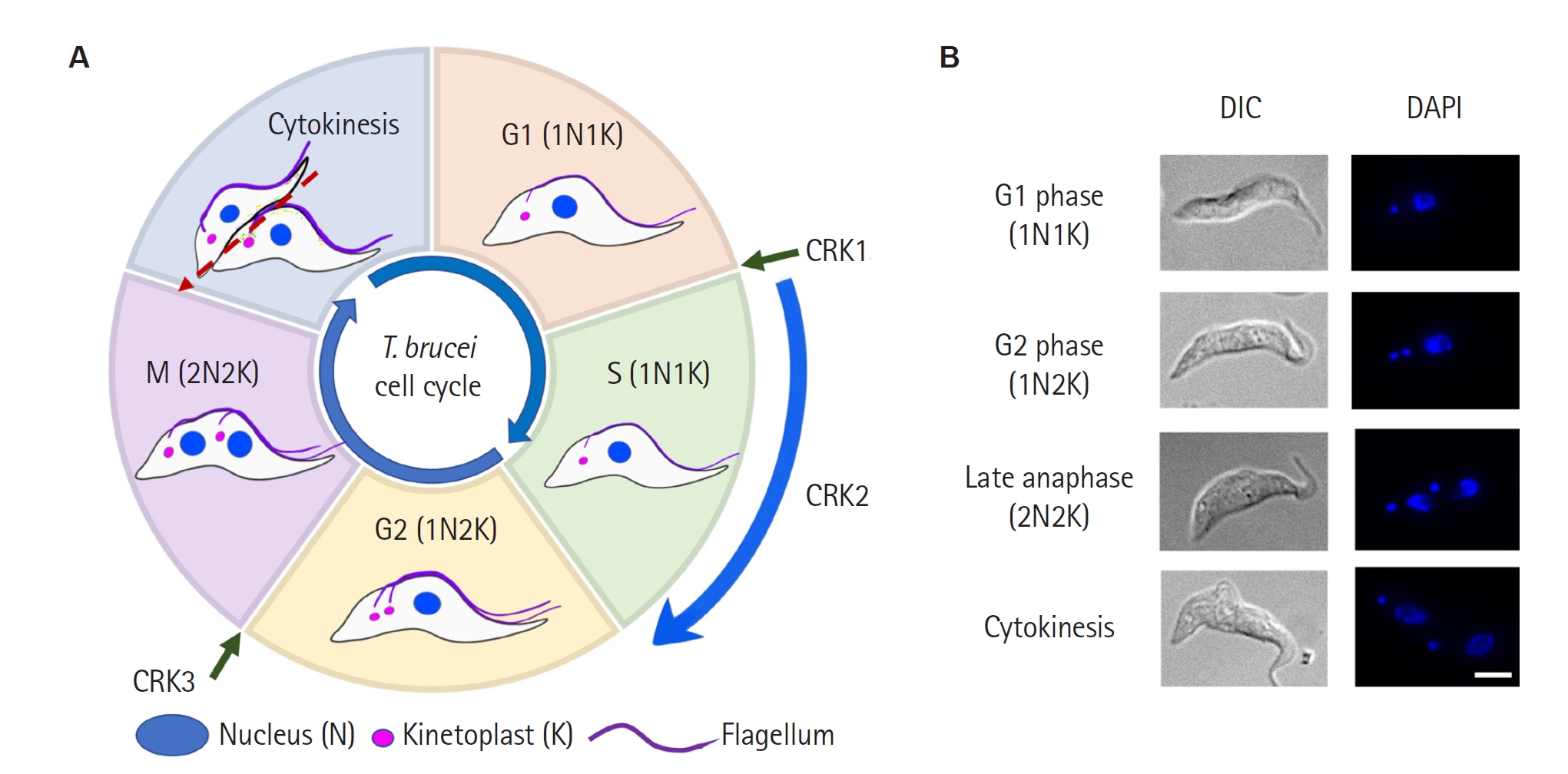
Fig. 2.Molecular mechanism by which CRK2–CYC13 promotes DNA replication initiation in Trypanosoma brucei. (A) Comparison of replication initiation in a canonical eukaryote and in T. brucei. In canonical systems, S-CDK phosphorylates Sld2/Sld3, which together with Dpb11 recruit GINS and Cdc45 to Mcm2–7 to assemble the active Cdc45–Mcm2–7–GINS (CMG) helicase, while Dbf4-dependent kinase (DDK) phosphorylates Mcm2–7. In T. brucei, Cdc6, Cdt1, Sld2, Sld3, and DDK are absent. Instead, CRK2–CYC13 directly phosphorylates Mcm3 at Thr-310. (B) Step-by-step mechanism. (1) CRK2–CYC13 recognizes Mcm2–7. (2) CRK2 phosphorylates Mcm3-Thr310. (3) Phospho-Mcm3 promotes binding of the GINS subunit Sld5. (4) Full assembly of an active CMG helicase, with Cdc45 and the remaining GINS subunits (Psf1, Psf2, and Psf3) joining without disruption of pre-existing Mcm3–Mcm2 or Mcm3–Cdc45 contacts.
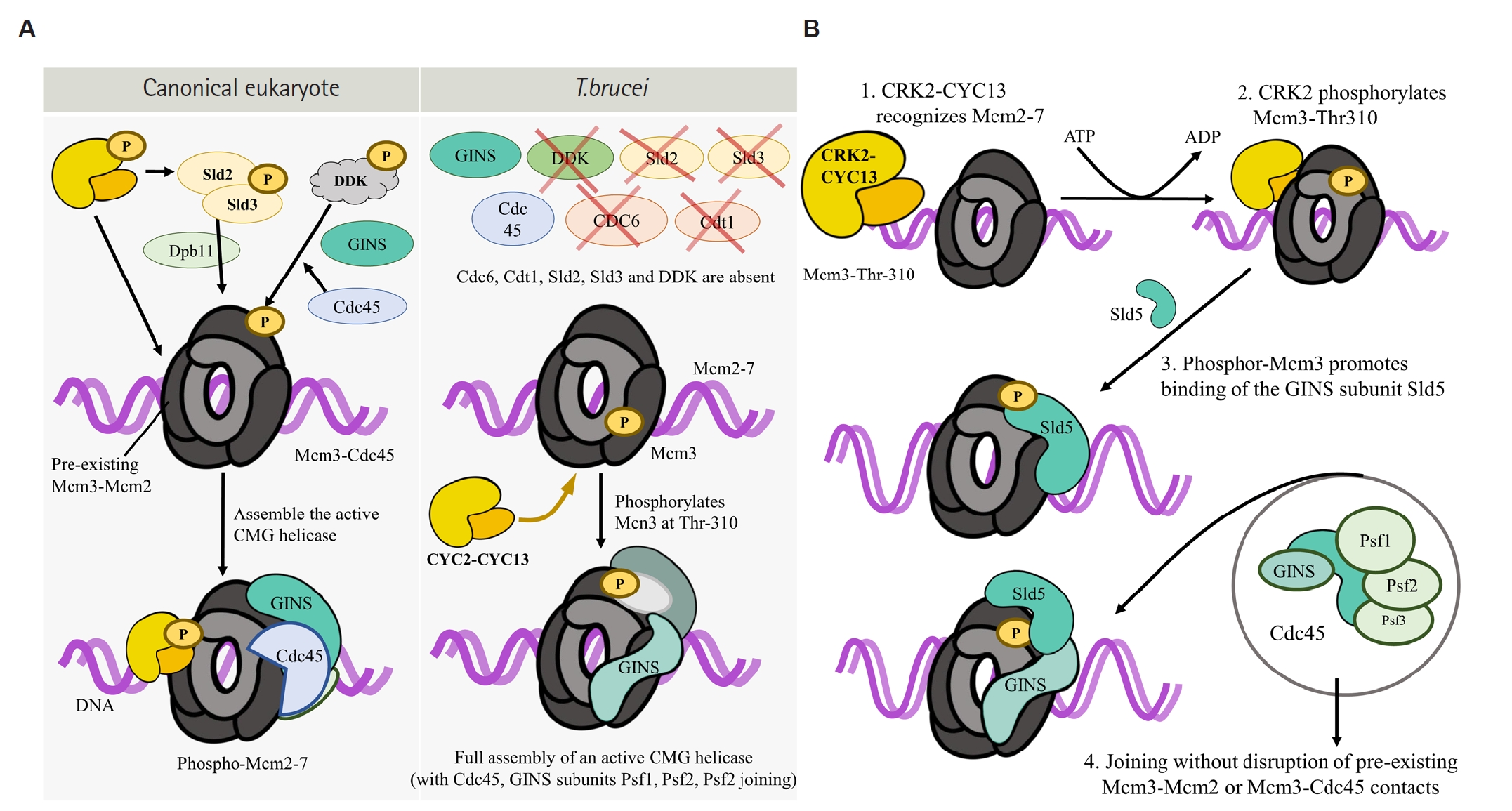
Fig. 3.Integrative map of Cdc2-related kinase (CRK)–cyclin modules in the Trypanosoma brucei cell cycle. The central wheel depicts the canonical cell-cycle phases (G1, S, G2, M, and cytokinesis). CRK1 (G1/S transition), CRK2–CYC13 (S phase), and CRK3 (G2/M transition) are positioned at their implicated phases; cytokinesis is regulated by non-CRK factors (TbPLK, TbAUK1, and CIF1–CIF4 network). Modules are color-coded by depth of mechanistic validation: validated CRK–cyclin–substrate axis (green, CRK2–CYC13–Mcm3); cyclin partner known with substrates proposed but in vivo complex unresolved (orange, CRK1); cyclin partner known with substrate only from in vitro assays (yellow, CRK3); essential CRK modules whose links to canonical cell-cycle transitions are indirect or incompletely resolved (gray, CRK9–CYC12–CRK9AP and CRK12–CYC9). CRK4 and CRK6 are omitted because functional cyclin partners and direct substrates have not been reproducibly established.
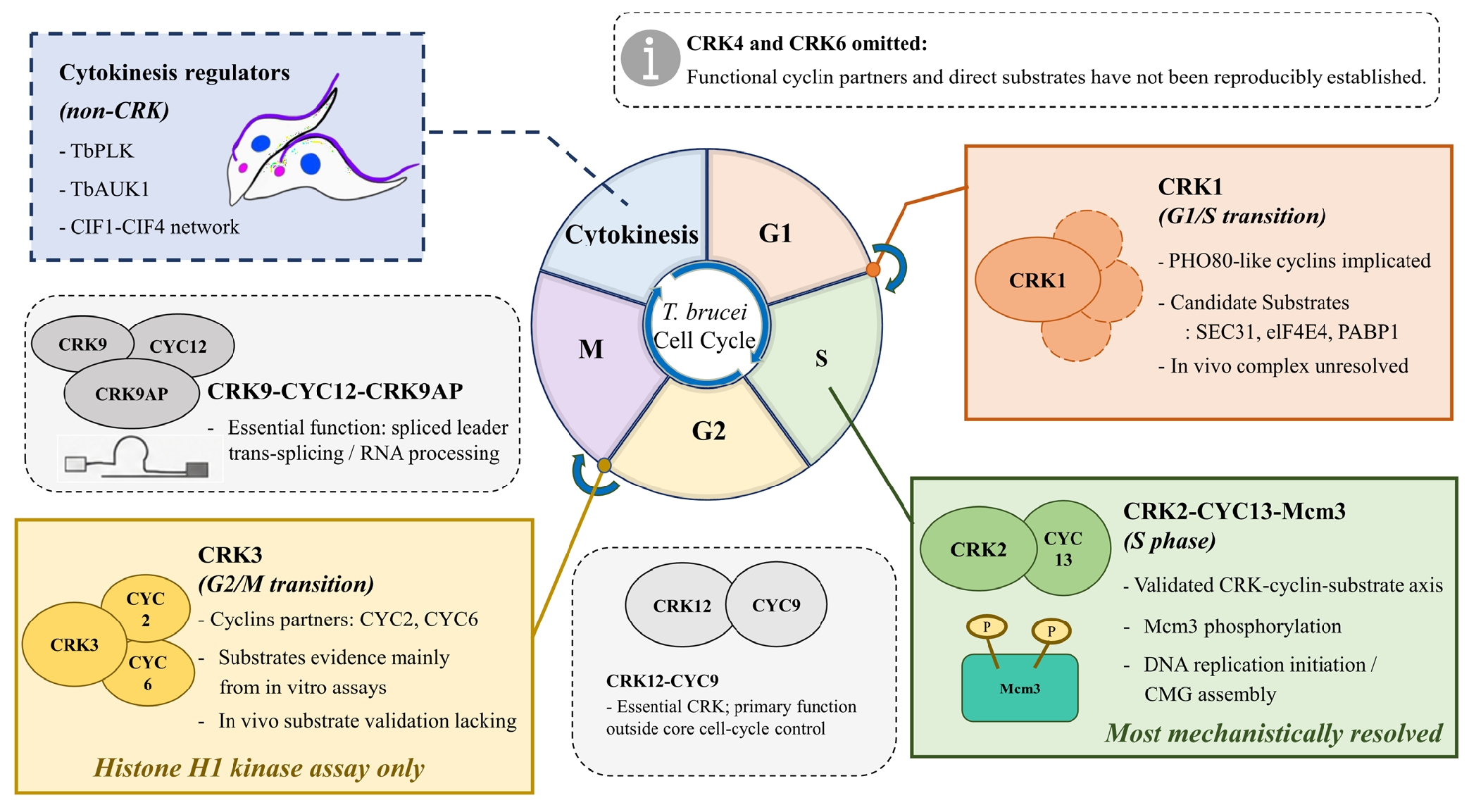
Table 1.Overview of CRK-associated regulatory modules, substrates, and unresolved questions in Trypanosoma brucei
Table 1.
|
CRK/module |
Reported/proposed partner |
Implicated process/stage |
Known or proposed substrate/target |
Representative reference |
Unresolved issue |
|
CRK1 |
PHO80-like cyclins, including CYC2/4/5/7 |
G1/S transition; posterior morphogenesis; anterograde protein transport; translation initiation |
Reported CRK1-phosphorylated targets: Sec31; eIF4E4; PABP1 |
[15,16,22-24] |
Cyclin-specific CRK1 complex composition and substrate specificity remain unclear |
|
CRK2 |
CYC2 |
G1/S-related association (proposed) |
Physiological substrate unclear |
[27] |
Whether CRK2–CYC2 forms a functional kinase complex in vivo remains unclear |
|
CYC13 |
S phase; DNA replication |
Mcm3 phosphorylation (Ser-213, Thr-310); Mcm3–Sld5 interaction/CMG assembly |
[17] |
In vivo substrate identity and cyclin-specific function remain unresolved |
|
CRK3 |
CYC2; CYC6 |
G2/M transition; mitotic progression |
Histone H1 used as assay substrate; physiological substrate unclear |
[16,21,22,26] |
In vivo substrate identity and exact cyclin specificity remain unresolved |
|
CRK9 |
CYC12; CRK9AP |
Pre-mRNA processing / transcription-coupled regulation; indirect cell-cycle effects |
RNA-processing machinery; direct cell-cycle substrate unclear |
[18,19,25] |
Whether cell-cycle phenotypes are direct or secondary remains unclear |
|
CRK12 |
CYC9 |
Essential CRK–cyclin kinase module; endocytosis and cytokinesis-related phenotypes |
Physiological substrate unclear |
[20] |
Direct cell-cycle substrate and mechanistic link between CRK12–CYC9 activity, endocytosis, and cytokinesis remain unclear |
References
- 1. World Health Organization. Trypanosomiasis, human African (sleeping sickness). World Health Organization; 2023.
- 2. Okello I, Mafie E, Eastwood G, Nzalawahe J, Mboera LEG. African animal trypanosomiasis: a systematic review on prevalence, risk factors and drug resistance in sub-Saharan Africa. J Med Entomol 2022;59:1099-143. https://doi.org/10.1093/jme/tjac018
- 3. World Health Organization. Guidelines for the treatment of human African trypanosomiasis. World Health Organization; 2024.
- 4. Lindner AK, Lejon V, Chappuis F, et al. New WHO guidelines for treatment of gambiense human African trypanosomiasis including fexinidazole: substantial changes for clinical practice. Lancet Infect Dis 2020;20:e38-46. https://doi.org/10.1016/S1473-3099(19)30612-7
- 5. Lindner AK, Lejon V, Barrett MP, et al. New WHO guidelines for treating rhodesiense human African trypanosomiasis: expanded indications for fexinidazole and pentamidine. Lancet Infect Dis 2025;25:e77-85. https://doi.org/10.1016/S1473-3099(24)00581-4
- 6. Gilbert IH. Drug discovery for neglected diseases: molecular target-based and phenotypic approaches. J Med Chem 2013;56:7719-26. https://doi.org/10.1021/jm400362b
- 7. Kubota K, Funabashi M, Ogura Y. Target deconvolution from phenotype-based drug discovery by using chemical proteomics approaches. Biochim Biophys Acta Proteins Proteom 2019;1867:22-7. https://doi.org/10.1016/j.bbapap.2018.08.002
- 8. La Greca F, Magez S. Vaccination against trypanosomiasis: can it be done or is the trypanosome truly the ultimate immune destroyer and escape artist? Hum Vaccin 2011;7:1225-33. https://doi.org/10.4161/hv.7.11.18203
- 9. Sherwin T, Gull K. The cell division cycle of Trypanosoma brucei brucei: timing of event markers and cytoskeletal modulations. Philos Trans R Soc Lond B Biol Sci 1989;323:573-88. https://doi.org/10.1098/rstb.1989.0037
- 10. Gluenz E, Povelones ML, Englund PT, Gull K. The kinetoplast duplication cycle in Trypanosoma brucei is orchestrated by cytoskeleton-mediated cell morphogenesis. Mol Cell Biol 2011;31:1012-21. https://doi.org/10.1128/MCB.01176-10
- 11. Li Z. Regulation of the cell division cycle in Trypanosoma brucei. Eukaryot Cell 2012;11:1180-90. https://doi.org/10.1128/EC.00145-12
- 12. Wheeler RJ, Gull K, Sunter JD. Coordination of the cell cycle in trypanosomes. Annu Rev Microbiol 2019;73:133-54. https://doi.org/10.1146/annurev-micro-020518-115617
- 13. Berriman M, Ghedin E, Hertz-Fowler C, et al. The genome of the African trypanosome Trypanosoma brucei. Science 2005;309:416-22. https://doi.org/10.1126/science.1112642
- 14. Hammarton TC. Cell cycle regulation in Trypanosoma brucei. Mol Biochem Parasitol 2007;153:1-8. https://doi.org/10.1016/j.molbiopara.2007.01.017
- 15. Liu Y, Hu H, Li Z. The cooperative roles of PHO80-like cyclins in regulating the G1/S transition and posterior cytoskeletal morphogenesis in Trypanosoma brucei. Mol Microbiol 2013;90:130-46. https://doi.org/10.1111/mmi.12352
- 16. Tu X, Wang CC. The involvement of two cdc2-related kinases (CRKs) in Trypanosoma brucei cell cycle regulation and the distinctive stage-specific phenotypes caused by CRK3 depletion. J Biol Chem 2004;279:20519-28. https://doi.org/10.1074/jbc.M312862200
- 17. Lee KJ, Li Z. The CRK2-CYC13 complex functions as an S-phase cyclin-dependent kinase to promote DNA replication in Trypanosoma brucei. BMC Biol 2021;19:29. https://doi.org/10.1186/s12915-021-00961-1
- 18. Badjatia N, Park SH, Ambrósio DL, Kirkham JK, Günzl A. Cyclin-dependent kinase CRK9, required for spliced leader trans splicing of pre-mRNA in trypanosomes, functions in a complex with a new L-type cyclin and a kinetoplastid-specific protein. PLoS Pathog 2016;12:e1005498. https://doi.org/10.1371/journal.ppat.1005498
- 19. Gosavi U, Srivastava A, Badjatia N, Günzl A. Rapid block of pre-mRNA splicing by chemical inhibition of analog-sensitive CRK9 in Trypanosoma brucei. Mol Microbiol 2020;113:1225-39. https://doi.org/10.1111/mmi.14489
- 20. Monnerat S, Almeida Costa CI, Forkert AC, et al. Identification and functional characterisation of CRK12:CYC9, a novel cyclin-dependent kinase (CDK)-cyclin complex in Trypanosoma brucei. PLoS One 2013;8:e67327. https://doi.org/10.1371/journal.pone.0067327
- 21. Hammarton TC, Clark J, Douglas F, Boshart M, Mottram JC. Stage-specific differences in cell cycle control in Trypanosoma brucei revealed by RNA interference of a mitotic cyclin. J Biol Chem 2003;278:22877-86. https://doi.org/10.1074/jbc.M300813200
- 22. Tu X, Wang CC. Pairwise knockdowns of cdc2-related kinases (CRKs) in Trypanosoma brucei identified the CRKs for G1/S and G2/M transitions and demonstrated distinctive cytokinetic regulations between two developmental stages of the organism. Eukaryot Cell 2005;4:755-64. https://doi.org/10.1128/EC.4.4.755-764.2005
- 23. Hu H, Gourguechon S, Wang CC, Li Z. The G1 cyclin-dependent kinase CRK1 in Trypanosoma brucei regulates anterograde protein transport by phosphorylating the COPII subunit Sec31. J Biol Chem 2016;291:15527-39. https://doi.org/10.1074/jbc.M116.715185
- 24. An T, Liu Y, Gourguechon S, Wang CC, Li Z. CDK phosphorylation of translation initiation factors couples protein translation with cell-cycle transition. Cell Rep 2018;25:3204-14.e5. https://doi.org/10.1016/j.celrep.2018.11.063
- 25. Gourguechon S, Wang CC. CRK9 contributes to regulation of mitosis and cytokinesis in the procyclic form of Trypanosoma brucei. BMC Cell Biol 2009;10:68. https://doi.org/10.1186/1471-2121-10-68
- 26. Van Hellemond JJ, Neuville P, Schwarz RT, Matthews KR, Mottram JC. Isolation of Trypanosoma brucei CYC2 and CYC3 cyclin genes by rescue of a yeast G(1) cyclin mutant. Functional characterization of CYC2. J Biol Chem 2000;275:8315-23. https://doi.org/10.1074/jbc.275.12.8315
- 27. Gourguechon S, Savich JM, Wang CC. The multiple roles of cyclin E1 in controlling cell cycle progression and cellular morphology of Trypanosoma brucei. J Mol Biol 2007;368:939-50. https://doi.org/10.1016/j.jmb.2007.02.050
- 28. McKean PG. Coordination of cell cycle and cytokinesis in Trypanosoma brucei. Curr Opin Microbiol 2003;6:600-7. https://doi.org/10.1016/j.mib.2003.10.010
- 29. Zhou Q, Gu J, Lun ZR, Ayala FJ, Li Z. Two distinct cytokinesis pathways drive trypanosome cell division initiation from opposite cell ends. Proc Natl Acad Sci U S A 2016;113:3287-92. https://doi.org/10.1073/pnas.1601596113
- 30. Bell SP, Dutta A. DNA replication in eukaryotic cells. Annu Rev Biochem 2002;71:333-74. https://doi.org/10.1146/annurev.biochem.71.110601.135425
- 31. Bleichert F, Botchan MR, Berger JM. Mechanisms for initiating cellular DNA replication. Science 2017;355:eaah6317. https://doi.org/10.1126/science.aah6317
- 32. Tu X, Mancuso J, Cande WZ, Wang CC. Distinct cytoskeletal modulation and regulation of G1-S transition in the two life stages of Trypanosoma brucei. J Cell Sci 2005;118:4353-64. https://doi.org/10.1242/jcs.02567
- 33. Tu X, Wang CC. Coupling of posterior cytoskeletal morphogenesis to the G1/S transition in the Trypanosoma brucei cell cycle. Mol Biol Cell 2005;16:97-105. https://doi.org/10.1091/mbc.e04-05-0368
- 34. da Silva MS, Pavani RS, Damasceno JD, et al. Nuclear DNA replication in trypanosomatids: there are no easy methods for solving difficult problems. Trends Parasitol 2017;33:858-74. https://doi.org/10.1016/j.pt.2017.08.002
- 35. Marques CA, Tiengwe C, Lemgruber L, et al. Diverged composition and regulation of the Trypanosoma brucei origin recognition complex that mediates DNA replication initiation. Nucleic Acids Res 2016;44:4763-84. https://doi.org/10.1093/nar/gkw147
- 36. Dang HQ, Li Z. The Cdc45·Mcm2-7·GINS protein complex in trypanosomes regulates DNA replication and interacts with two Orc1-like proteins in the origin recognition complex. J Biol Chem 2011;286:32424-35. https://doi.org/10.1074/jbc.M111.240143
- 37. Tiengwe C, Marcello L, Farr H, et al. Identification of ORC1/CDC6-interacting factors in Trypanosoma brucei reveals critical features of origin recognition complex architecture. PLoS One 2012;7:e32674. https://doi.org/10.1371/journal.pone.0032674
- 38. Walker RG, Thomson G, Malone K, et al. High throughput screens yield small molecule inhibitors of Leishmania CRK3:CYC6 cyclin-dependent kinase. PLoS Negl Trop Dis 2011;5:e1033. https://doi.org/10.1371/journal.pntd.0001033
- 39. Cayla M, Nievas YR, Matthews KR, Mottram JC. Distinguishing functions of trypanosomatid protein kinases. Trends Parasitol 2022;38:950-61. https://doi.org/10.1016/j.pt.2022.08.009
- 40. Zhou Q, Hu H, Li Z. An EF-hand-containing protein in Trypanosoma brucei regulates cytokinesis initiation by maintaining the stability of the cytokinesis initiation factor CIF1. J Biol Chem 2016;291:14395-409. https://doi.org/10.1074/jbc.M116.726133
- 41. Kurasawa Y, Hu H, Zhou Q, Li Z. The trypanosome-specific protein CIF3 cooperates with the CIF1 protein to promote cytokinesis in Trypanosoma brucei. J Biol Chem 2018;293:10275-86. https://doi.org/10.1074/jbc.RA118.003113
- 42. Hu H, An T, Kurasawa Y, Zhou Q, Li Z. The trypanosome-specific proteins FPRC and CIF4 regulate cytokinesis initiation by recruiting CIF1 to the cytokinesis initiation site. J Biol Chem 2019;294:16672-83. https://doi.org/10.1074/jbc.RA119.010538