Abstract
This study presents 2 cases of wound myiasis in a dog and a cat involving morphological and molecular identification in Korea. A 7-year-old male Jindo mix dog, Canis familiaris, was admitted to a veterinary clinic in Yangsan-si, Gyeongsangnam-do, Korea, in July 2024. The bite wound near the right rib was infected with maggots. We carefully removed the 5 maggots using forceps. The wound healed without any subsequent complications. A Korean Shorthair stray cat, Felis catus, was brought to the same clinic in October 2024 after being hit by a car. The maggot-infected wounds were found in the anus and perianal regions. We removed all 10 maggots from the wounds. After treatment, no living maggots were observed; however, the cat was euthanized due to trauma-related complications. The maggots from the dog and the cat were third instars with 12 body segments and posterior spiracles containing 3 distinct slits. Compared with the dog-derived larvae, the cat-derived larvae had a more dorsally positioned posterior process of the mouth hook, reduced dorsal papillae distances, and distinctly sclerotized ventral cornua. Complete cox1 gene analysis confirmed the dog- and cat-derived larvae as Lucilia sericata (Meigen, 1826) and Lucilia illustris (Meigen, 1826), respectively, whereas 18S rRNA gene analysis showed limited species-level resolution. In the present study, wound myiasis caused by L. sericata and L. illustris is described in a dog and a cat in Korea based on morphological and molecular findings.
-
Key words: Myiasis, larva, Calliphoridae, dogs, cats
Introduction
Myiasis is the infection of vertebrates, including humans and animals, by larvae of flies that feed on living or dead tissue of their hosts [
1]. Anatomically, myiasis is classified based on the site of infection, including auricular, cutaneous, gastrointestinal, ophthalmic, oral, and urogenital forms [
2]. Ecologically, myiasis is categorized as specific (obligatory), semi-specific (facultative), or accidental (pseudomyiasis), depending on the degree to which the larvae rely on the host for survival and development [
3].
The most commonly observed form is cutaneous myiasis, which is generally subdivided into furuncular, migratory, and wound myiasis [
4]. Wound myiasis occurs when dipteran larvae infect open wounds in a living host. The most common flies that cause obligatory wound myiasis globally are
Cochliomyia hominivorax (Coquerel, 1858),
Chrysomya bezziana Villeneuve, 1914, and
Wohlfahrtia magnifica (Schiner, 1862) [
3], while facultative wound myiasis has been associated with the Muscidae, Calliphoridae, and Sarcophagidae families [
4]. Wound myiasis can cause tissue damage, delayed wound healing, and secondary infections, particularly in debilitated, injured, or outdoor animals, highlighting its clinical relevance in veterinary medicine [
3,
5].
Several cases of human myiasis have been reported in Korea, including nasal, submandibular, internal, and oral myiasis [
6-
9]. In contrast, myiasis in companion animals has rarely been documented, with only 2 canine cases reported to date, including cutaneous myiasis associated with tick infestation and wound myiasis caused by
Lucilia sericata (Meigen, 1826) [
10,
11]. Therefore, species-level information on companion animal myiasis remains limited in Korea, and feline wound myiasis has not been well documented. The present study aimed to report wound myiasis in a domestic dog and a stray cat and to identify the causative fly species using morphological and molecular analyses.
Case Report
Case 1
A 7-year-old male Jindo mix dog,
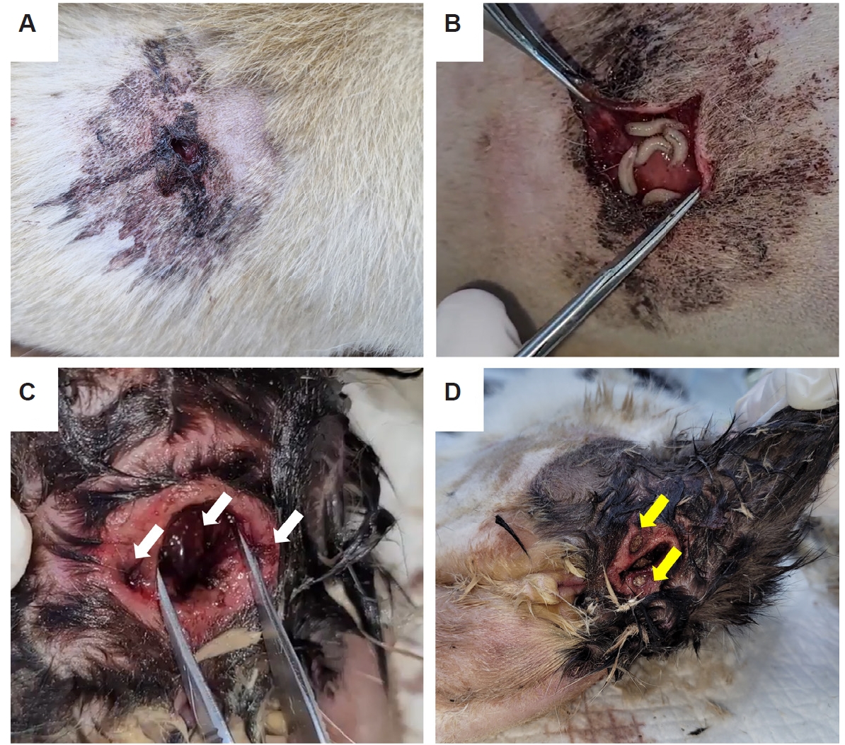
Canis familiaris, raised outdoors, was bitten by a stray dog in July 2024. This study was approved by the Institutional Animal Care and Use Committee of Chungbuk National University (CBNUA-26-0064-03). The dog was brought to a veterinary clinic in Yangsan-si (si means city), Gyeongsangnam-do (do means province), Korea, and the veterinarians found a wound near the right rib infected with dipteran larvae, leading to a diagnosis of wound myiasis (
Fig. 1A,
B). The dog showed lameness at presentation. Five maggots were carefully removed using forceps. A drain was inserted into the wound site to facilitate the drainage of exudates and inflammatory substances. At the 1-week follow-up, the drain was removed, and the wound healed completely without any subsequent complications.
In October 2024, a Korean shorthair stray cat,
Felis catus, was brought to the same veterinary clinic 4 days after being hit by a car. A physical examination revealed a wound in the anus and perianal region (
Fig. 1C,
D). The wound was filled with 10 maggots, which were removed using forceps. Necrotic tissue was removed and the wound was washed with 0.9% normal saline. Topical 1% ivermectin was applied to target maggots within necrotic tissue or inaccessible areas. Additionally, antibiotic treatment was administered. Although no maggots were observed after treatment, euthanasia was performed because of hind limb paralysis and complete loss of urination and defecation functions associated with a fractured fifth lumbar vertebra (L5).
All maggots collected from the domestic dog and stray cat were preserved in 70% ethanol and transferred to the Veterinary Parasitology Laboratory, College of Veterinary Medicine, Chungbuk National University, Korea, for species identification. The remaining larvae were preserved and retained as specimens in the same laboratory. For morphological examination, representative well-preserved larvae appropriate for microscopic observation were selected from each case.
Two larvae from the dog and 3 larvae from the cat were examined under a light microscope. The examined larvae were consistent with third-instar larvae based on posterior spiracular morphology, with 3 distinct slits observed in each spiracle [
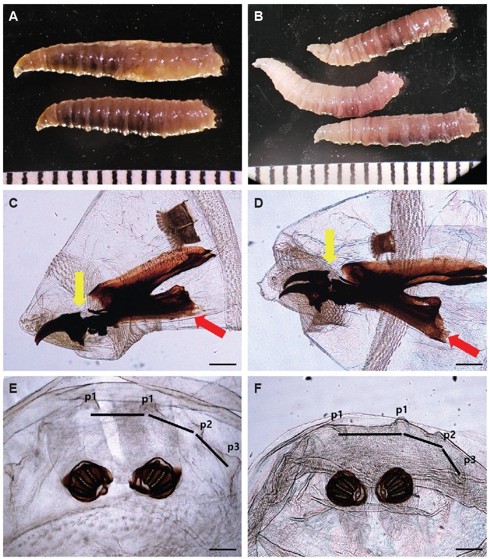
12]. The larvae from the dog measured approximately 13 mm in length and 2 mm in width, whereas those from the cat measured approximately 12 mm in length and 2 mm in width. In both hosts, the larval body was whitish-beige, and reddish internal contents were visible through the cuticle. The larvae were subcylindrical, slightly tapered anteriorly, composed of 12 segments, and the posterior margins of each segment were covered with numerous single-pointed spines. General external morphology appeared similar between the 2 groups.
Species identification was based on cephaloskeletal and posterior papillae characteristics described in published diagnostic keys for third-instar
Lucilia larvae [
12,
13]. The posterior process of the mouth hook was positioned more dorsally in the cat-derived larvae than in the dog-derived larvae. The distance between the median dorsal papillae (p1–p1) and between adjacent dorsal papillae (p1–p2 and p2–p3) was almost uniform around the posterior spiracle in the dog-derived larvae. In contrast, these distances were noticeably reduced in the cat-derived larvae. Additionally, the posterior tip of the ventral cornua was poorly sclerotized or indistinct in the dog-derived larvae but distinctly sclerotized in the cat-derived larvae. These morphological differences supported the identification of the dog-derived larvae as
L. sericata and the cat-derived larvae as
Lucilia illustris (Meigen, 1826) (
Fig. 2A-
F). However, because the number of diagnostic characters available in larval specimens is limited and closely related
Lucilia species can be difficult to distinguish morphologically, representative larvae from each case were further subjected to molecular analysis for species confirmation.
For molecular identification, DNA was extracted from a single maggot from a domestic dog and a stray cat, respectively. Genomic DNA was extracted using the DNeasy Blood & Tissue Kit (Qiagen), following the manufacturer's instructions. DNA concentrations were confirmed using a spectrophotometer (DeNovix). PCR was conducted to amplify the complete cytochrome c oxidase subunit 1 (
cox1) gene of each species of maggot using 2 primer sets (
Table 1) [
14-
16]. Since amplifying the full-length
cox1 gene in a single PCR was unsuccessful, 2 partially overlapping fragments were amplified and assembled to obtain the complete sequence. The PCR products were sent to Macrogen for direct sequencing. Additionally, the complete 18S rRNA gene was amplified using a primer set common to eukaryotes (
Table 1) [
14-
16]. The PCR products were subsequently cloned into
Escherichia coli DH5α using the pGEM-T easy vector (Promega). For each sample, at least 3 colonies were selected and sequenced using a universal primer set (M13F and M13R) by Macrogen.
The obtained sequences were aligned using MEGA version 11, and species identification was performed through the BLAST (
https://blast.ncbi.nlm.nih.gov/Blast.cgi), and phylogenetic analysis using MEGA version 11 with the maximum likelihood method. The bootstrap method with 500 replicates was used to assess the reliability of the tree.
The complete
cox1 gene (1,539 bp) of each maggot species was successfully sequenced and served as the primary marker for species-level confirmation. The sequences obtained from the maggots collected from the domestic dog and those from the stray cat showed 94.5% interspecies identity. The
L. sericata sequence showed 99.9% identity with GenBank sequences from New Zealand (AJ417713) and Australia (MW255540). The
L. illustris sequence, obtained from a maggot collected from the stray cat, showed 99.8% identity with
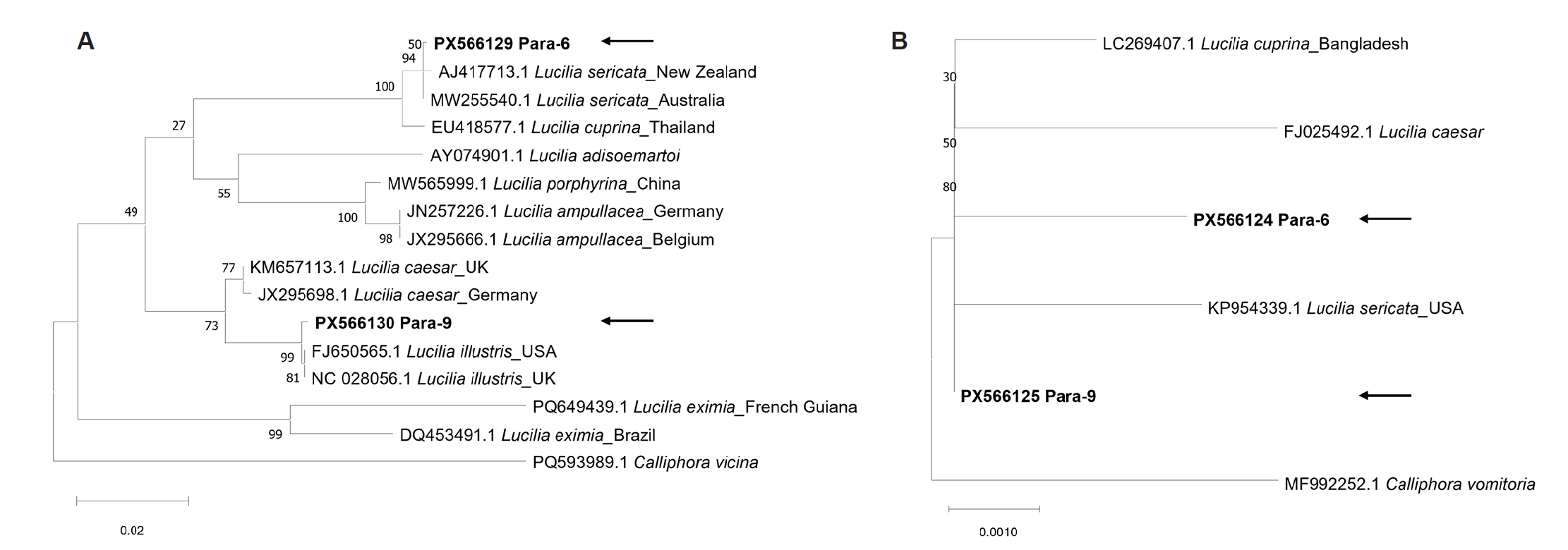
L. illustris (FJ650565 and NC028056) deposited in the GenBank database. The phylogenetic analysis of
cox1 revealed a clear separation of
L. sericata and
L. illustris (
Fig. 3A).
The complete 18S rRNA gene (1,984 bp) was also sequenced to provide additional molecular data for species identification. However, 18S rRNA analysis showed limited resolution for species-level differentiation. The 18S rRNA sequences of
L. sericata and
L. illustris showed 99.8% interspecies identity. The
L. sericata sequence obtained in this study showed 99.9% identity with
L. sericata (KP954339) deposited in the GenBank database. However, the identity of the
L. illustris sequence could not be confirmed due to the absence of a reference sequence in the GenBank database. Unlike the
cox1 analysis, the 18S rRNA gene phylogenetic analysis could not determine the precise species due to the lack of a
Lucilia reference database (
Fig. 3B).
All sequences obtained in this study were deposited in the GenBank database (18S rRNA gene, PX566124-PX566125; cox1, PX566129-PX566130).
Discussion
Although both
L. sericata and
L. illustris are commonly found in Korea, maggot infections caused by these species, referred to as myiasis, have rarely been reported in companion animals [
11]. This may be because veterinary clinicians primarily focus on wound treatment and may not fully recognize the importance of case reporting and species identification. Unfortunately, maggot samples recovered from clinical cases are often discarded without being transferred from clinical settings to a parasitology laboratory. Nevertheless, accurately identifying the species of fly is crucial because myiasis caused by certain species can lead to severe or malignant cases. Traditionally, fly species identification relied on morphological analysis under a microscope. However, identifying morphologically similar species can be challenging. For instance,
L. illustris and
Lucilia caesar (Linnaeus, 1758) are nearly identical, and males are the only stage that can be reliably distinguished. Additionally, species identification can be achieved by rearing the larvae to the adult stage and examining their morphological characteristics. However, this approach is time-consuming and requires expertise and optimal environmental conditions [
11].
To address these challenges, molecular methods have become more common. These approaches offer faster, more reliable results, even with immature or dead specimens. Recent studies have focused on various molecular markers, including mitochondrial and nuclear genes, to differentiate between closely related species and improve the accuracy of identification in the forensic, medical, and veterinary fields [
11]. In the present study, a molecular analysis of the complete
cox1 gene revealed that the maggots from a domestic dog and a stray cat were
L. sericata and
L. illustris, respectively. However, the species could not be determined using the 18S rRNA gene due to the limited reference sequences for the
Lucilia genus in the database.
L. sericata, commonly known as the green bottle fly, lays eggs on necrotic tissue, wounded skin, or diseased areas, where the larvae primarily feed [
2]. This species is one of the most widespread causative agents of myiasis worldwide and has been reported to infect various hosts, including cats, dogs, and humans [
11]. Canine myiasis caused by
L. sericata has been reported in Italy [
1], Turkiye [
17], Iran [
18], Israel [
19], and Korea [
10,
11]. In the present case, species identification of canine myiasis was confirmed to be
L. sericata.
L. illustris is an important agent of wound myiasis in humans and livestock. It is often found in association with wounds, feces, and carcasses. This species is considered one of the primary contributors to sheep blowfly strike. Previous studies have reported
L. illustris in association with cat carcasses; however, to our knowledge, no published clinical case of wound myiasis caused by this species in a living cat has been reported [
20]. Therefore, the present case may represent the first report of feline wound myiasis caused by
L. illustris.
These Lucilia species have been reported as causative agents of human myiasis. Therefore, the identification of these species in a dog and a cat suggests the public health relevance of Lucilia-associated myiasis in environments shared by humans and animals, where both may be exposed to the same flies. These findings highlight the importance of monitoring myiasis in stray and outdoor animals.
Both cases exhibited predisposing conditions that likely facilitated fly infection. Various predisposing factors increase the risk of myiasis in animals, including poor hygiene, traumatic injuries, dermatological conditions, and physical impairments that limit mobility or grooming behavior [
5]. In particular, wounded or debilitated animals raised outdoors are at substantially higher risk of fly infection [
10]. In case 1, the dog was raised outdoors and had an open wound, conditions that likely increased its exposure to flies and facilitated larval infection. Furthermore, this occurred during the summer when fly activity is high. The stray cat reported in case 2 was a free-roaming animal, a lifestyle that makes it susceptible to wounds from fights, car accidents, animal bites, or falls [
1]. These environmental and behavioral factors can make animals more vulnerable to myiasis.
This study has several limitations. Only 2 clinical cases were examined, and the larvae were not reared to adult flies for adult-stage morphological confirmation. In addition, molecular analysis was performed on one representative larva from each case rather than on all recovered larvae; therefore, mixed-species infection cannot be completely excluded. However, the cox1-based molecular identification was consistent with the morphological analysis of the examined representative larvae. Furthermore, 18S rRNA gene analysis had limited resolution for species-level differentiation because of the limited availability of Lucilia reference sequences in GenBank. Therefore, additional case reports and expanded reference sequence data are needed to better understand companion animal myiasis caused by Lucilia spp. in Korea.
This study provides the case-based evidences of canine and feline wound myiasis caused by Lucilia spp. in Korea. To our knowledge, this is the first reported case of wound myiasis associated with L. illustris in a living cat. The causative fly species were identified through both larval morphological examination and complete cox1 gene analysis, highlighting the importance of combining these approaches for accurate identification of Lucilia species. This report may serve as a reference for the diagnosis and species-level identification of Lucilia-associated wound myiasis in dogs and cats.
Notes
-
Data availability
The sequence data were deposited in the NCBI GenBank database (18S rRNA gene, PX566124–PX566125; cox1, PX566129–PX566130). Specimens collected during this study will be made available to qualified researchers upon reasonable request.
-
Author contributions
Conceptualization: Lee SH. Data curation: Lee S, Lee SH. Investigation: Lee S, Kim J, Yang K, Choe S. Methodology: Lee S, Lee SH. Resources: Kim J, Yang K. Supervision: Lee SH. Validation: Lee S, Choe S, Lee SH. Visualization: Lee S. Writing – original draft: Lee S. Writing – review & editing: Lee S, Kim J, Yang K, Choe S, Lee SH.
-
Conflict of interest
Seung-Hun Lee serves as an editor of Parasites, Hosts and Diseases but had no involvement in the decision to publish this article. No other potential conflicts of interest relevant to this study were reported.
Fig. 1.Gross findings of wound myiasis in a domestic dog and a stray cat. A bitten scar (A) on the right rib area of a dog and maggots (B) in the opened wound. A wound (C) in the anal and perianal regions of a stray cat, indicated by white arrows, and dipteran larvae (D) on the affected region, indicated by yellow arrows.
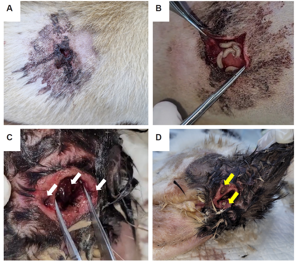
Fig. 2.Maggots of (A) Lucilia sericata and (B) Lucilia illustris were recovered from the wounds in the respective cases. The cephaloskeletons of (C) L. sericata and (D) L. illustris: the posterior process of the mouth hook (yellow arrow) was more dorsally positioned in L. illustris than in L. sericata, and the posterior tip of the ventral cornua (red arrow) was indistinct in L. sericata but distinctly sclerotized in L. illustris. The spiracular openings (posterior spiracles) of the 2 fly larvae species showed similar distances between papillae in (E) L. sericata, whereas the distances were unequal in (F) L. illustris. Scale bars: (A, B) 1 mm; (C-F) 200 μm.
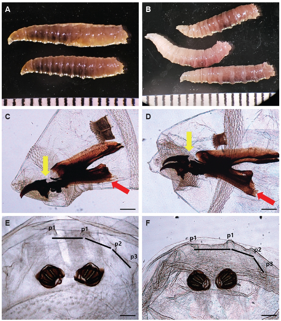
Fig. 3.Phylogenetic trees of Lucilia spp. Based on (A) cox1, (B) 18S rRNA gene. To construct the phylogenetic tree, the maximum likelihood method was used with 500 bootstrap replications. The sequences analyzed in this tree were obtained from the GenBank databases. The sequences identified in this study are indicated in bold and with arrows.
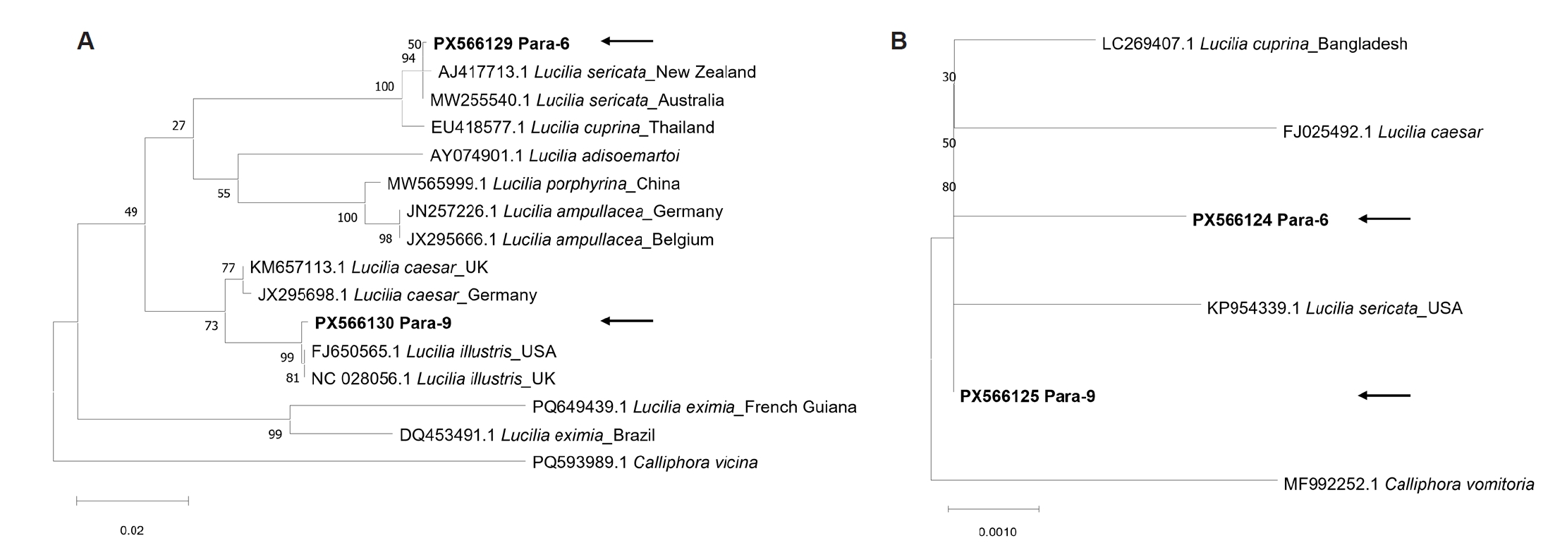
Table 1.Primers used to identify the maggot species from a domestic dog and a stray cat in Korea
Table 1.
|
Target gene |
Primers (5’ to 3’) |
PCR condition |
Expected size (bp) |
Reference |
|
cox1
|
CTGCTACTTTATGAGCTTTAGG |
94℃/5 min; 30 cycles: 94℃/30 sec, 55℃/30 sec, 72℃/1 min; 72℃/15 min |
560 |
[14,15]a
|
|
CCATTGCACTAATCTGCCA |
|
CCTTTAGAATTGCAGTCTAATGTCA |
95℃/11 min; 35 cycles: 95℃/30 sec, 55℃/30 sec, 72℃/1 min 30 sec; 72℃ 15 min |
1,400 |
[15] |
|
CAAGTTGTGTAAGCATC |
|
18S rRNA |
AACCTGGTTGATCCTGCCAGT |
95℃/2 min; 35 cycles: 92℃/30 sec, 55℃/30 sec, 72℃/45 sec, 72℃ 7 min |
1,900 |
[16] |
|
TGATCCTTCTGCAGGTTCACCTAC |
References
- 1. Pezzi M, Scapoli C, Chicca M, et al. Cutaneous myiasis in cats and dogs: cases, predisposing conditions and risk factors. Vet Med Sci 2021;7:378-84. https://doi.org/10.1002/vms3.370
- 2. Pezzi M, Bonacci T, Leis M, et al. Myiasis in domestic cats: a global review. Parasit Vectors 2019;12:372. https://doi.org/10.1186/s13071-019-3618-1
- 3. Francesconi F, Lupi O. Myiasis. Clin Microbiol Rev 2012;25:79-105. https://doi.org/10.1128/CMR.00010-11
- 4. McGraw TA, Turiansky GW. Cutaneous myiasis. J Am Acad Dermatol 2008;58:907-26. https://doi.org/10.1016/j.jaad.2008.03.014
- 5. Pezzi M, Whitmore D, Chicca M, Lanfredi M, Leis M. Traumatic myiasis caused by an association of Sarcophaga tibialis (Diptera: Sarcophagidae) and Lucilia sericata (Diptera: Calliphoridae) in a domestic cat in Italy. Korean J Parasitol 2015;53:471-5. https://doi.org/10.3347/kjp.2015.53.4.471
- 6. Kim JS, Seo PW, Kim JW, et al. A nasal myiasis in a 76-year-old female in Korea. Korean J Parasitol 2009;47:405-7. https://doi.org/10.3347/kjp.2009.47.4.405
- 7. Joo CY, Kim JB. Nosocomial submandibular infections with dipterous fly larvae. Korean J Parasitol 2001;39:255-60. https://doi.org/10.3347/kjp.2001.39.3.255
- 8. Chung PR, Jung Y, Kim KS, et al. A human case of internal myiasis in Korea. Korean J Parasitol 1996;34:151-4. https://doi.org/10.3347/kjp.1996.34.2.151
- 9. Jang M, Ryu SM, Kwon SC, et al. A case of oral myiasis caused by Lucilia sericata (Diptera: Calliphoridae) in Korea. Korean J Parasitol 2013;51:119-23. https://doi.org/10.3347/kjp.2013.51.1.119
- 10. Choi J, Kim H, Na J, Kim S, Park C. Cutaneous myiasis associated with tick infestations in a dog. J Vet Clin 2015;32:473-5. https://doi.org/10.17555/jvc.2015.10.32.5.473
- 11. Choe S, Lee D, Park H, et al. Canine wound myiasis caused by Lucilia sericata (Diptera: Calliphoridae) in Korea. Korean J Parasitol 2016;54:667-71. https://doi.org/10.3347/kjp.2016.54.5.667
- 12. Szpila K. Key for the identification of third instars of European blowflies (Diptera: Calliphoridae) of forensic importance. In: Amendt J, Goff ML, Campobasso CP, Grassberger M, editors. Current concepts in forensic entomology. Springer; 2009. p. 43-56.
- 13. Velásquez Y, Magaña C, Martínez‐Sánchez A, Rojo S. Diptera of forensic importance in the Iberian Peninsula: larval identification key. Med Vet Entomol 2010;24:293-308. https://doi.org/10.1111/j.1365-2915.2010.00879.x
- 14. Chen WY, Hung TH, Shiao SF. Molecular identification of forensically important blow fly species (Diptera: Calliphoridae) in Taiwan. J Med Entomol 2004;41:47-57. https://doi.org/10.1603/0022-2585-41.1.47
- 15. Park SH, Zhang Y, Piao H, et al. Use of cytochrome c oxidase subunit I (COI) nucleotide sequences for identification of the Korean Luciliinae fly species (Diptera: Calliphoridae) in forensic investigations. J Korean Med Sci 2009;24:1058-63. https://doi.org/10.3346/jkms.2009.24.6.1058
- 16. Mangold AJ, Bargues MD, Mas-Coma S. 18S rRNA gene sequences and phylogenetic relationships of European hard-tick species (Acari: Ixodidae). Parasitol Res 1998;84:31-7. https://doi.org/10.1007/s004360050352
- 17. Gökpinar S, Karsli B. Traumatic myiasis associated with Wohlfahrtia magnifica and Lucilia sericata larvae in dog. Van Vet J 2018;29:55-7.
- 18. Moshaverinia A, Mehrjerdi HK. Canine myiasis and its causal agents in Northeastern Iran. Iran J Parasitol 2016;11:91-7.
- 19. Schnur HJ, Zivotofsky D, Wilamowski A. Myiasis in domestic animals in Israel. Vet Parasitol 2009;161:352-5. https://doi.org/10.1016/j.vetpar.2009.01.026
- 20. Bagsby K, Hans K. Lucilia silvarum Meigen (Diptera: Calliphoridae) is a primary colonizer of domestic cats (Felis catus). Insects 2024;15:32. https://doi.org/10.3390/insects15010032