Abstract
Malaria is the leading cause of death and economic burden worldwide. Current diagnostic methods rely on microscopy and blood-based rapid diagnostic tests. A common rapid diagnostic test target is Plasmodium falciparum histidine-rich protein 2 (PfHRP2), a parasite protein released into the host's blood during Plasmodium infection; it has also been detected in saliva, although results have been inconsistent. This pilot study quantified plasma and salivary PfHRP2 antigen levels and assessed their associations with age, parasite density, and clinical malaria status in a malaria-endemic region of western Kenya with high transmission. This pilot study enrolled 34 patients with acute uncomplicated malaria. Paired blood and saliva samples were collected at enrolment, with repeat sampling 4 weeks after treatment. P. falciparum infection was confirmed by light microscopy, and PfHRP2 concentrations were measured using an enzyme-linked immunosorbent assay. Data were analyzed in R version 4.5.2. Spearman’s rank correlation and robust linear regression were used to examine the relationship between plasma and salivary P. falciparum antigen levels in individuals with acute malaria and to assess whether PfHRP2 concentrations in either matrix predicted parasite density, clinical malaria status, and age. Salivary PfHRP2 levels did not significantly predict parasite density or age (χ²=0.54, P=0.76). By contrast, plasma PfHRP2 levels strongly predicted both parasite density and age (P=0.034). Plasma and salivary PfHRP2 concentrations were not significantly correlated (ρ=-0.327, P=0.059). Nevertheless, routine measurement of salivary PfHRP2 using more sensitive methods may improve diagnosis and guide treatment management.
-
Key words: Rapid diagnostic test, malaria, microscopy, enzyme-linked immunosorbent assay, Saliva
Introduction
Malaria remains a major public health concern and a leading cause of morbidity and mortality in the tropics [
1]. An estimated 247 million malaria cases occurred worldwide in 2021 across 84 malaria-endemic countries, up from 245 million in 2020; countries in the World Health Organization African Region accounted for most of this increase [
2]. In 2021, more than 40% of global investment in malaria was channeled through the Global Fund to Fight AIDS, Tuberculosis and Malaria. Countries and partners raised a total of 15.9 billion US dollars, the largest replenishment in the fund’s history [
2]. However, malaria elimination efforts continue to face setbacks associated with the coronavirus disease 2019 pandemic and treatment and control campaigns, among other challenges. Improved malaria diagnosis is therefore urgently needed to reduce malaria-related deaths and economic burden. Timely, accurate diagnosis followed by effective treatment is the primary strategy for malaria treatment [
3].
Microscopy measures peripheral parasitemia and remains the gold standard for malaria diagnosis [
4], although it has limitations [
5] and is less reliable at low parasite levels [
6]. Malaria rapid diagnostic tests (RDTs) detect
Plasmodium-specific proteins, such as
Plasmodium falciparum histidine-rich protein 2 (PfHRP2), in infected blood [
7]. PfHRP2-based tests have demonstrated accuracy in detecting
P. falciparum infection [
8]. However, both methods require blood collection, which is labor-intensive and time-consuming and requires considerable expertise and trained healthcare workers [
9]. These invasive procedures also cause discomfort and pain [
10]. Saliva has shown considerable potential as a noninvasive specimen for detecting malaria antigens, although findings have been mixed [
11]. Among children with malaria in Accra, Ghana, detection of PfHRP2 in whole saliva had a sensitivity of 43% and a specificity of 53% [
12]. Another study using a blood-based RDT detected
P. falciparum lactate dehydrogenase (PfLDH) with sensitivities of 77.9% in whole saliva and 96.7% in saliva supernatant [
4]. A study in Lagos State reported sensitivities for PfHRP2 detection of 96.7% in blood and 12.7% in saliva [
13]. Most studies of saliva for malaria diagnosis have been qualitative and have not quantified salivary antigens, making it difficult to determine whether low sensitivity reflects low antigen concentrations. The only study to quantify salivary PfHRP2 levels also found a good correlation between blood and salivary levels [
11]. However, that study was conducted in an area of low malaria endemicity and included relatively few participants. Whether similar patterns occur in a malaria-endemic region therefore requires investigation. The utility of salivary PfHRP2 levels as a proxy for
Plasmodium infection also needs to be evaluated. This pilot study detected and quantified PfHRP2 antigens in blood and saliva and examined their relationships with age, parasite density, and clinical malaria status in a highly malaria-endemic area of western Kenya.
Methods
Ethical statement
The School of Graduate Studies at Maseno University authorized this study. Ethical approval was obtained from the Maseno University Ethical Review Committee (MSU/DRPI/MUERC/00816/19). Participants’ rights to informed consent, privacy, and confidentiality were upheld throughout and after the study. Consent for participants younger than 18 years was sought from their parents or guardians. This observational study did not pose a potential for harm, damage, or injury; accordingly, a trained, qualified phlebotomist collected the blood samples. Participants received no compensation, but the study findings benefited the wider community.
Study site
The study was conducted at Chulaimbo Sub-County Hospital in Kisumu County, a malaria-endemic area of western Kenya [
14]. Residents receive approximately 100–300 infectious mosquito bites annually, and
P. falciparum causes 97% of malaria infections [
15]. According to the 2020 Kenya Malaria Indicator Survey, malaria prevalence among children aged 6 months to 14 years in Kisumu County was approximately 15.6%–19%. The hospital’s catchment area is 1,131 m above sea level and receives 1,200–1,300 mm of rainfall annually. Humidity ranges from 50 to 68 g/m³, and temperatures range from 20°C to 35°C [
14]. In 2021, the population of the Chulaimbo Hospital catchment area was approximately 21,452. The hospital predominantly serves the rural population of North West Kisumu ward, whose economic activities include small-scale farming and business [
14].
The study combined cross-sectional and longitudinal designs. Patients presenting to Chulaimbo Sub-County Hospital in Kisumu West Sub-County, Kenya, with symptoms of uncomplicated malaria were enrolled after consent was obtained. Blood and saliva were collected at enrollment and again from the same participants 4 weeks after treatment. Suspected clinical malaria was defined according to Kenya Ministry of Health guidelines.
The sample size for this pilot study was based on the statistical power needed to detect a moderate-to-strong correlation between systemic and oral malaria antigens. With a two-sided significance level (α) of 0.05 and target power (1−β) of 80%, the minimum sample size (n) was calculated using the formula for correlation coefficients:
where n is the minimum sample size required to detect the target effect size; Zα is the standard normal critical value for a 95% confidence interval (1.96); Zβ is the standard normal value for 80% statistical power (0.841); and C is Fisher’s Z-transformation of the target correlation coefficient (r=0.465).
Calculation:
A minimum of 34 patients (28 children and 6 adults) was therefore required to provide sufficient power to detect a moderate-to-strong diagnostic relationship between plasma and salivary PfHRP2 levels. This sample size permitted quantitative evaluation of antigen levels and assessment of their diagnostic utility in a highly malaria-endemic area of western Kenya.
Inclusion and exclusion criteria
Patients were eligible if they were at least 1 year old, had malaria symptoms, and had microscopically confirmed P. falciparum infection. Informed consent was also required: adults consented for themselves, and parents or guardians consented on behalf of children. Patients were excluded if they were not Kisumu County residents, had symptoms of an illness other than malaria, or had symptoms of severe malaria. Blood and saliva were collected from 34 participants (28 children and 6 adults) to quantify P. falciparum antigens. Matched blood and saliva samples from each participant were processed at Chulaimbo Sub-County Hospital. Hemoglobin concentrations were measured using the HemoCue method.
Blood sample collection and processing
A trained phlebotomist collected approximately 1 ml of whole blood from each participant by finger prick into EDTA-treated anticoagulant tubes. Samples were labeled immediately, placed in a cool box, and transported to the laboratory within 2 h of collection. At the laboratory, blood samples were centrifuged at 2,600 × g for 15 min (Eppendorf). Supernatants were transferred to fresh 2 ml tubes and stored at -20°C for batch analysis. Malaria was diagnosed by both light microscopy and RDTs, as shown in
Fig. 1.
Saliva was collected from each participant with confirmed
P. falciparum infection as previously described [
11]. Approximately 3 ml was collected into sterile cryovial tubes, immediately placed in a cool box maintained at 4°C, and transported to the laboratory within 2 h. On arrival, samples were centrifuged at 2,600 × g and 4°C for 15 min, as previously described [
11]. After collection, the following protease inhibitors were added to the saliva: aprotinin (Sigma-Aldrich, lot A1153; 0.9 μl, 6.12 units/ml), sodium orthovanadate (Sigma-Aldrich, S6508; 3 μl at 400 mM, pH 10 in water), and phenylmethylsulfonyl fluoride (My Biosphere, catalog No. 545833; 10 μl at 10 mg/ml in isopropyl alcohol). Samples were then stored at -20°C for batch analysis.
PfHRP2 levels in saliva and blood were quantified using a sandwich ELISA, following a previously described method [
11] with minor modifications. Immulon 4 HBX microtiter plates were coated with a PfHRP2 capture antibody (MyBioSource, catalog No. MBS532523) diluted in phosphate-buffered saline (PBS) to a final concentration of 0.1625 μg/ml, then incubated overnight at 4°C. The wells were washed 3 times with PBS containing 0.05% Tween-20 (PBS-T) and blocked for 1 h at room temperature with 4% (w/v) nonfat dry milk powder (Sigma-Aldrich) in PBS. After blocking, the plates were washed 3 times with PBS-T. Standards were prepared by diluting recombinant PfHRP2 in blocking buffer to an initial concentration of 5 ng/μl, followed by 1:1 serial dilutions in the same buffer for a total of 8 dilution steps. Standards, samples, and blanks were added in duplicate, and the plates were sealed with adhesive film and incubated for 1 h at room temperature. The plates were then emptied and washed 5 times with PBS-T. Biotinylated PfHRP2 detection antibody (MyBioSource, catalog No. 6120632), diluted in blocking buffer to a final concentration of 125 ng/ml, was added, followed by incubation for 1 h at room temperature. The plates were washed 5 times with PBS-T, and peroxidase-labeled streptavidin (0.8 μg/ml; Thermo Fisher Scientific, catalog No. 21126) was added. After incubation for 30 min at room temperature with gentle agitation, the plates were washed 5 times with PBS-T. A chemiluminescent substrate (Thermo Fisher Scientific, catalog No. 37070) was then added, and the plates were incubated for 25 min at room temperature. Luminescence was measured immediately at 460/20 nm in relative light units using a Synergy HTX multimode reader (Agilent Technologies/BioTek). Sample PfHRP2 concentrations were extrapolated from a standard curve generated from known recombinant PfHRP2 concentrations.
Data were analyzed in R version 4.5.2. P. falciparum antigen levels in saliva and blood were measured using a highly sensitive multimode ELISA reader. To address the inherent right-skewness of biomarker concentrations, all antigen levels (plasma and salivary PfHRP2) and parasite density values were log10-transformed before analysis, stabilizing variance across measurement scales. Robust linear regression related log10-transformed antigen levels to parasite density, clinical malaria status, and age. Logistic regression was used to model clinical malaria as a binary outcome (1=clinical malaria, 0=post-treatment follow-up). Clinical malaria was defined according to Kenya Ministry of Health guidelines for suspected uncomplicated malaria: an axillary temperature >37.0°C plus at least 1 of chills, headache, or severe malaise, together with microscopic confirmation of P. falciparum monoinfection. Spearman’s rank correlation was used to assess the correlation between blood and salivary PfHRP2 levels. Chi-square tests assessed associations between categorical variables, including salivary PfHRP2 detectability (detectable versus nondeductible) and clinical malaria status. A two-sided P<0.05 was considered statistically significant for all tests.
Results
Demographic characteristics of study participants
Table 1 presents participant demographics and laboratory findings during acute uncomplicated malaria and 4 weeks after treatment. Thirty-four participants (28 children and 6 adults) who presented to Chulaimbo Hospital with clinical malaria symptoms were enrolled after consent was obtained. Most participants were female (18/34, 53%). Mean weight (range) was 18.7 (11–28.8) kg in children and 59.9 (51.8–75.8) kg in adults. At enrollment, median age (interquartile range) was 6.6 (4.4) years in children and 26.4 (11.2) years in adults. Median parasite density (interquartile range) was higher in children, at 20,000 (21,400), than in adults, at 6,230 (9,370) (
Table 1).
Relationships of histidine-rich protein 2 levels with Plasmodium parasite density and age
To evaluate salivary PfHRP2 as a proxy for
Plasmodium infection, robust linear regression was used to examine the relationships of ELISA-measured PfHRP2 levels with blood smear parasite density and age. Salivary PfHRP2 levels did not significantly predict parasite density or age in individuals with acute malaria (
χ²=0.54,
P=0.76). The negative coefficients for log parasite density (β=-0.1384,
t=-0.7314) and age (β=-0.0037,
t=-0.3939) further suggested an inverse, although nonsignificant, relationship between systemic parasite load across ages and salivary antigen concentration (
Table 2). By contrast, plasma PfHRP2 levels strongly predicted parasite density and age in patients with acute malaria (
P=0.034), with a positive coefficient for log parasite density (β=0.2211,
t=1.2470) and a negative coefficient for age (β=-0.0435,
t=-4.919) (
Table 2).
Robust linear regression was used to assess the relationship between clinical malaria and antigen levels. Plasma PfHRP2 showed a strong positive trend in predicting clinical malaria status (odds ratio, 8.73; 95% confidence interval, 0.620–623.42), whereas salivary PfHRP2 had a low odds ratio (0.23; 95% confidence interval, 0.003–3.99) (
Table 3).
The correlation between plasma and salivary PfHRP2 levels in individuals with acute malaria was assessed to determine whether saliva could serve as a non-invasive diagnostic alternative to plasma. Salivary PfHRP2 did not show a positive linear relationship with either systemic antigen levels (ρ=-0.327,
P=0.059) (
Fig. 2) or total parasite burden (ρ=-0.246,
P=0.161) (
Table 4).
Discussion
Table 1 presents participant demographics and laboratory findings during acute uncomplicated malaria and 4 weeks after treatment. Microscopy provided estimates of total parasite burden with greater precision and sensitivity. This overall assessment indicated high parasite density after treatment, particularly in children (
Table 1). This finding may reflect markers associated with resistance to artemisinin combination therapy within a genetically heterogeneous parasite population [
16], together with lower population immunity to clear parasites, DNA, and malaria antigens [
17]. A subpopulation of dormant parasites is also the most likely explanation for delayed parasite clearance. During artemisinin-induced dormancy, a subset of asexual parasites temporarily enters a state of arrested growth and metabolic inactivity, allowing survival during artemisinin treatment [
18]. These dormant parasites may be killed slowly by partner drugs while contributing to persistent parasite detection after treatment.
The utility of salivary PfHRP2 levels as a proxy for
Plasmodium infection was evaluated using ELISA measurements. Relationships with blood smear parasite density, clinical malaria, and age were examined. The bivariate correlation between log plasma PfHRP2 concentration and log parasite density (
Table 2), together with the positive relationship between plasma PfHRP2 and clinical malaria (
Table 3), indicates a significant association between elevated plasma levels and symptomatic illness. These findings agree with those of Manning et al. [
19] in Papua New Guinea and Martiáñez-Vendrell [
20]. However, salivary PfHRP2 levels did not predict parasite density in individuals with acute malaria. The odds ratio for salivary PfHRP2 was also lower than that for plasma PfHRP2, suggesting that the salivary test produced a markedly weaker signal than blood-based measurements despite confirmed clinical malaria. This may primarily reflect the lower ELISA detection limit for antigens in saliva than in plasma observed in the results above. Protein transport into saliva depends on molecular mass, ionization, and salivary ph. Molecules may be diluted to different extents during transfer from plasma to saliva [
11].
P. falciparum antigens also differ in stability between saliva and plasma. Salivary proteases may break down PfHRP2 [
21], further reducing the sensitivity of salivary malaria-antigen detection. Saliva clarity varies among individuals even when patients are asked to provide unstimulated drool. Obtaining clear samples was particularly difficult in dehydrated children. Taken together, the findings indicate substantial decoupling of systemic and oral biomarkers. Plasma PfHRP2 remains a gold-standard indicator of malaria infection, whereas salivary PfHRP2 appears subject to distinct biological constraints. The lack of a strong predictive relationship suggests that standard salivary ELISA cannot yet directly replace blood-based testing without accounting for oral clearance or degradation. Future research should therefore prioritize substantially more sensitive salivary diagnostic tools or robust standardization methods that correct for oral matrix effects.
Age significantly predicted median plasma PfHRP2 levels but not salivary levels. Its highly significant contribution indicated that younger patients tended to have much higher systemic antigen levels. This finding is consistent with the high-transmission setting of western Kenya, where older individuals develop partial immunity and clear antigens more efficiently [
22]. Previous studies similarly reported that parasite density declines with age [
23], peaking in young children and decreasing thereafter [
24]. Malaria antigen levels also increase with parasite density [
25]. Median PfHRP2 levels were therefore expected to be higher in children than in adults in both plasma and saliva, but this pattern was observed only in plasma. Direct age-stratified studies of salivary PfHRP2 are scarce, and most data come from small cohorts. For example, a 2012 pilot study reported levels <200 pg/ml in adolescents aged 13–15 years and up to 1,167 pg/ml in adults aged 23–57 years [
11]. Research on plasma PfHRP2 nevertheless provides a robust explanation, because transfer into saliva depends on the plasma load. The longer PfHRP2 half-life in semi-immune adults results in antigen accumulation owing to mature immune modulation and renal/hepatic function [
26]. Children, however, clear the antigen more rapidly through innate responses [
27].
This study examined the relationship between plasma and salivary PfHRP2 levels in patients with malaria to determine whether saliva could replace plasma as a diagnostic specimen. The negative correlation suggests that detectable salivary levels tend to decrease as systemic antigen concentrations increase, with the relationship approaching statistical significance. Parasite density likewise showed a weak, nonsignificant negative correlation with salivary PfHRP2. Together, these findings provide empirical evidence of decoupling between salivary antigen levels and systemic parasite biomass. This divergence may reflect low assay sensitivity and inhibitors in saliva [
21]. PfHRP2 is also less stable in saliva than in plasma. Compared with plasma, saliva contains proteases that may break down the enzymes [
11], potentially reducing salivary
P. falciparum antigen levels. In this study, however, the ELISA protocol was optimized for the salivary matrix by using a highly sensitive multimode reader capable of detecting luminescence. This significantly improved sensitivity, quantification, and overall reliability compared with standard colorimetric methods. Protease inhibitors were also added to saliva to reduce protease activity. When added immediately after collection, these inhibitors suppress enzymatic activity by binding competitively or reversibly to proteolytic enzymes’ active sites, arresting catalysis and preserving antigen integrity. To further preserve sample integrity, saliva was collected by passive drooling. Samples were collected consistently during the morning to minimize circadian effects on flow rate. Participants were also instructed to refrain from eating, drinking, or performing oral hygiene for 30–60 min before collection to reduce matrix effects and contamination. Samples were collected at 4°C and transported to the laboratory within 2 h for processing to limit post collection variability. Nevertheless, variation in sample clarity, particularly in dehydrated children despite requests for unstimulated drool, may have affected antigen detection and consistency. This may explain the lack of correlation between plasma and salivary antigen levels in individuals with acute malaria.
Fig. 2 illustrates the negative relationship between log-transformed plasma and salivary PfHRP2 concentrations in the 34 patients with acute malaria. The age color gradient indicates that this pattern persisted across the demographic range, from children to adults. This negative association supports the hypothesis that complex biological barriers in the oral cavity, such as antigen degradation by salivary proteases [
11], may decouple salivary PfHRP2 levels from systemic parasite burden. Future studies should standardize saliva-collection protocols and ensure appropriate processing before analysis to improve salivary data.
In contrast to these findings, a study in Msambweni, Kwale County, found a slight positive linear relationship between blood and salivary PfLDH levels [
28]. That study included 175 participants, compared with 34 in the present study, which may explain the slight positive relationship between plasma and salivary antigen levels. Future studies should increase the sample size to improve statistical power and detect subtle correlations, because larger cohorts have shown better proportionality for antigens such as PfLDH [
21]. Differences between the target antigens, PfLDH and PfHRP2, may also reflect fundamental differences in their biochemical properties, secretion patterns, and behaviour in fluids such as saliva. PfHRP2 persists for weeks after treatment because of slow clearance, with a plasma half-life of approximately 7–14 days [
11]. This persistence reflects cumulative parasite biomass rather than active infection. Salivary concentrations, however, are typically low (e.g., 17–1,167 pg/ml), and detection often correlates poorly with concurrent blood parasitemia. Salivary PfHRP2 may therefore primarily reflect accumulated or historical biomass rather than real-time active replication, consistent with its role as an indicator of exposure intensity or cumulative burden in low-density or post-treatment settings rather than acute parasitemia. PfLDH is a soluble glycolytic enzyme produced by viable parasites and is rapidly cleared from circulation after parasite death, making it a better marker of active infection in saliva [
21]. In larger cohorts, PfLDH also appears to be more proportional to blood parasitemia, although this relationship remains weak. This may reflect differences in molecular size, approximately 140 kDa [
21]. Both antigens, however, present challenges for salivary detection, including potential degradation by proteases, inhibitors, and matrix effects.
Saliva is a promising diagnostic fluid for malaria when protein degradation and matrix effects are reduced, but the present study found no statistically significant relationship between median plasma and salivary PfHRP2 levels under the experimental conditions used. A more appropriate approach to malaria diagnosis would combine a test kit sufficiently sensitive to detect low salivary antigen levels with standardized sample collection and processing. Investigating other malaria antigens present in both plasma and saliva may yield different results and could form the basis of future work.
This study found no statistically significant association between plasma and salivary PfHRP2 levels in individuals with acute malaria. Nevertheless, routine measurement of salivary PfHRP2 using more sensitive methods may provide clinicians and researchers with a more robust, real-time dataset that could improve diagnosis and guide treatment management.
Notes
-
Data availability
The data are available from the authors upon request.
-
Author contributions
Conceptualization: Omingo RA, Ayieko C. Data curation: Omingo RA, Ayieko C, Onyango PO. Formal analysis: Omingo RA, Ayieko C, Onyango PO. Funding acquisition: Ayieko C. Investigation: Omingo RA, Ayieko C, Onyango PO. Methodology: Omingo RA, Ayieko C, Onyango PO. Project administration: Ayieko C. Resources: Omingo RA, Ayieko C, Onyango PO, Sati BO, Omwenga VK, Ogolla S. Software: Omingo RA, Ayieko C, Onyango PO, Sati BO. Supervision: Ayieko C, Onyango PO, Ogolla S. Validation: Ayieko C, Onyango PO, Sati BO, Omwenga VK, Ogolla S. Visualization: Omingo RA, Ayieko C. Writing – original draft: Omingo RA. Writing – review & editing: Ayieko C.
-
Conflict of interest
The authors have no conflicts of interest to declare.
-
Funding
This study was funded by the Wellcome Trust through an HRCS Postdoctoral Grant (reference 209329/Z/17/Z).
-
Acknowledgments
The authors thank the study participants, guardians, and parents who provided samples. We are grateful to the Kenya Medical Research Institute-Kisian for permitting sample processing and storage at its facility. We also thank Mr. Ben Okeyo Sati, Boniface Ariera, Kevin Waomba, and Emilly Koech for helping collect samples at Chulaimbo Sub-County Hospital. Gertrude Wekesa and Regina Njeri of the International Livestock Research Institute provided guidance on sample analysis by ELISA at their facility. We thank Mr. Philip N. Ochien’g, Walter Odipo, Barnabas Muya, Joab Pira, and Julius Adundo, laboratory technicians in the Department of Zoology at Maseno University, for their technical assistance and guidance during the laboratory experiments.
Fig. 1.Malaria diagnosis by microscopy and rapid diagnostic test (RDT).
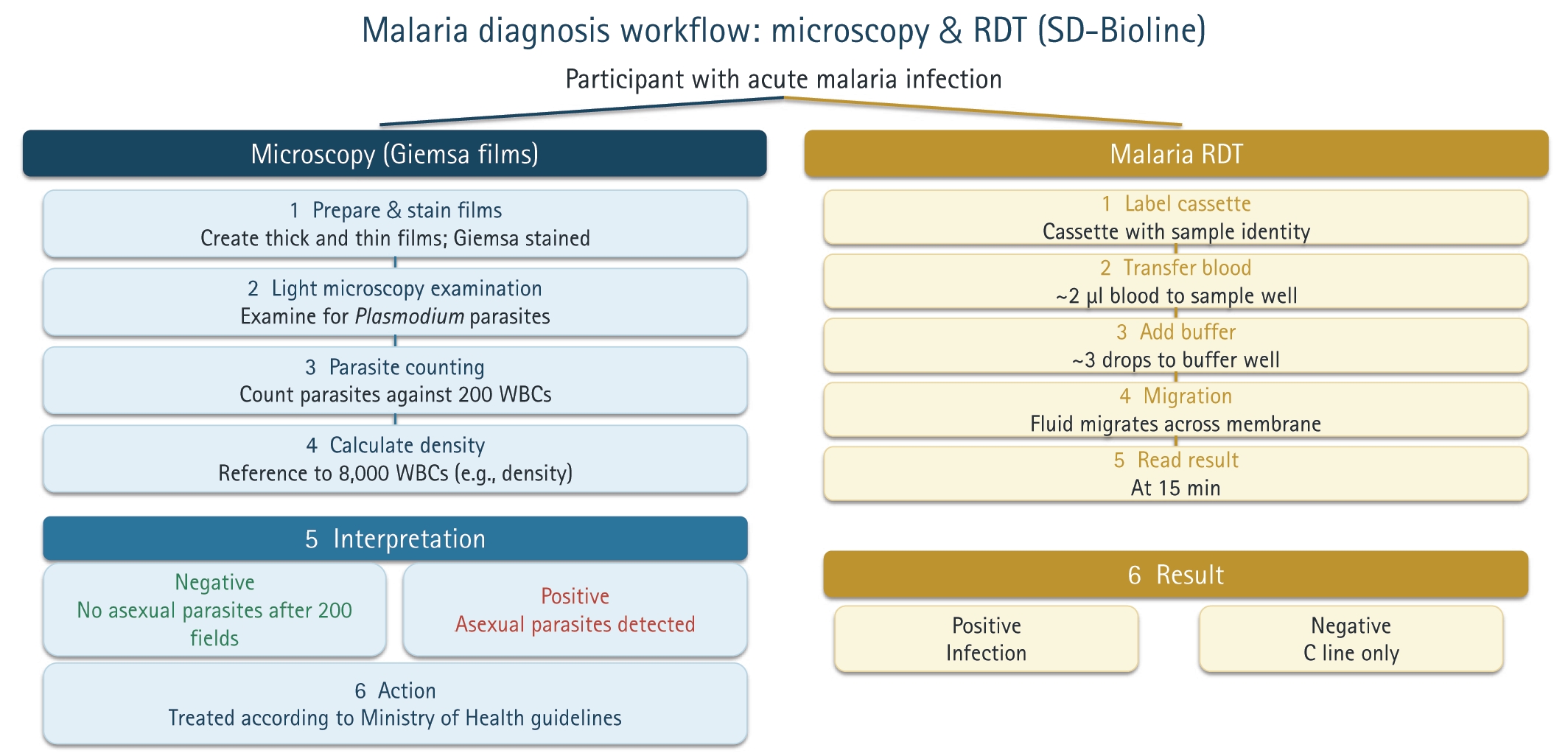
Fig. 2.Correlation between salivary and plasma histidine-rich protein 2 levels in children and adults with acute malaria. The plot shows the relationship between log-transformed plasma and salivary Plasmodium falciparum histidine-rich protein 2 (PfHRP2) concentrations in 34 patients with acute malaria. The analysis indicated a marginal inverse correlation (ρ=−0.327, P=0.059), shown by the negative slope of the regression line and the shaded 95% confidence interval. This downward trend suggests that higher plasma antigen levels do not correspond to higher salivary concentrations; the divergence between the 2 compartments approaches statistical significance. The age color gradient indicates that this pattern persists across the demographic range, from children to adults. This negative association supports the hypothesis that complex biological barriers in the oral cavity, including antigen degradation by salivary proteases or a saturation effect at the blood–saliva barrier, may decouple salivary PfHRP2 levels from systemic parasite burden.
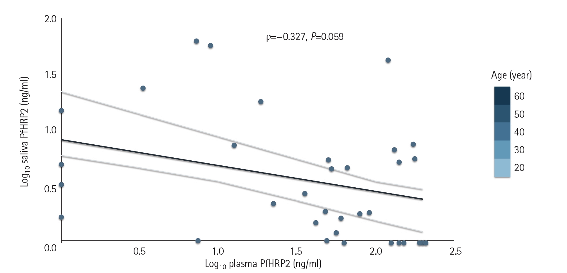
Table 1.Age and clinical characteristics of study participants
Table 1.
|
Children (n=28) |
Adults (n=6) |
|
Acute |
Post-treatment |
P-valuea
|
Acute |
Post-treatment |
P-valuea
|
|
Age (year) |
6.6 (4.4) |
6.6 (4.4) |
1.000 |
26.4 (11.2) |
26.4 (11.2) |
1.000 |
|
Hemoglobin (g/dl) |
11.1 (2.4) |
11.9 (1.3) |
0.236 |
14.9 (1.2) |
15.2 (0.7) |
0.317 |
|
Parasite density (/μl) |
20,000 (21,400) |
13,200 (0) |
<0.001 |
6,230 (9,370) |
0 (0) |
0.068 |
|
Temperature (°C) |
38.4 (1.0) |
36.4 (0.5) |
<0.001 |
36.4 (1.6) |
35.7 (0.4) |
0.109 |
Table 2.Robust linear regression analysis of factors influencing plasma and salivary histidine-rich protein 2 concentrations (n=34)
Table 2.
|
Predictor variable |
Predicted variable |
Coefficient (β) |
Standard error |
t-value |
|
Plasma PfHRP2a
|
log10(parasite density) |
0.2211 |
0.1773 |
1.2470 |
|
Age (year) |
-0.0435 |
0.0088 |
-4.9199 |
|
Salivary PfHRP2b
|
log10(parasite density) |
-0.1384 |
0.1893 |
-0.7314 |
|
Age (year) |
-0.0037 |
0.0094 |
-0.3939 |
Table 3.Logistic regression analysis predicting clinical malaria status
Table 3.
|
Predictor |
Odds ratio |
95% CI (lower bound) |
95% CI (upper bound) |
|
log10(plasma PfHRP2) |
8.7339 |
0.6203 |
623.428 |
|
log10(salivary PfHRP2) |
0.2362 |
0.0038 |
3.993 |
|
Age (year) |
0.8776 |
0.6971 |
0.9853 |
Table 4.Spearman’s rank correlation analysis of salivary histidine-rich protein 2 (n=34)
Table 4.
|
Correlation pair |
Spearman’s rho (ρ) |
P-value |
|
Plasma PfHRP2 vs. salivary PfHRP2 |
-0.3272 |
0.0589 |
|
Parasite density vs. salivary PfHRP2 |
-0.2459 |
0.1610 |
References
- 1. Okoyo C, Githinji E, Muia RW, et al. Assessment of malaria infection among pregnant women and children below five years of age attending rural health facilities of Kenya: a cross-sectional survey in two counties of Kenya. PLoS One 2021;16:e0257276. https://doi.org/10.1371/journal.pone.0257276
- 2. World Health Organization. World malaria report. World Health Organization; 2022.
- 3. Otambo WO, Olumeh JO, Ochwedo KO, et al. Health care provider practices in diagnosis and treatment of malaria in rural communities in Kisumu County, Kenya. Malar J 2022;21:129. https://doi.org/10.1186/s12936-022-04156-z
- 4. Nantavisai K. Malaria detection using non-blood samples. Songklanakarin J Sci Technol 2014;36:633-41.
- 5. Kifude CM, Rajasekariah HG, Sullivan DJ Jr, et al. Enzyme-linked immunosorbent assay for detection of Plasmodium falciparum histidine-rich protein 2 in blood, plasma, and serum. Clin Vaccine Immunol 2008;15:1012-8. https://doi.org/10.1128/CVI.00385-07
- 6. Berzosa P, de Lucio A, Romay-Barja M, et al. Comparison of three diagnostic methods (microscopy, RDT, and PCR) for the detection of malaria parasites in representative samples from Equatorial Guinea. Malar J 2018;17:333. https://doi.org/10.1186/s12936-018-2481-4
- 7. Amoah LE, Abankwa J, Oppong A. Plasmodium falciparum histidine rich protein-2 diversity and the implications for PfHRP 2: based malaria rapid diagnostic tests in Ghana. Malar J 2016;15:101. https://doi.org/10.1186/s12936-016-1159-z
- 8. Ly AB, Tall A, Perry R, et al. Use of HRP-2-based rapid diagnostic test for Plasmodium falciparum malaria: assessing accuracy and cost-effectiveness in the villages of Dielmo and Ndiop, Senegal. Malar J 2010;9:153. https://doi.org/10.1186/1475-2875-9-153
- 9. Tangpukdee N, Duangdee C, Wilairatana P, Krudsood S. Malaria diagnosis: a brief review. Korean J Parasitol 2009;47:93-102. https://doi.org/10.3347/kjp.2009.47.2.93
- 10. Owusu EDA, Campillo A, Daily J, Ding XC. Acceptance and perceived value of non-invasive malaria diagnostic tests in malaria-endemic countries. Malar J 2021;20:379. https://doi.org/10.1186/s12936-021-03911-y
- 11. Fung AO, Damoiseaux R, Grundeen S, et al. Quantitative detection of PfHRP2 in saliva of malaria patients in the Philippines. Malar J 2012;11:175. https://doi.org/10.1186/1475-2875-11-175
- 12. Wilson NO, Adjei AA, Anderson W, Baidoo S, Stiles JK. Detection of Plasmodium falciparum histidine-rich protein II in saliva of malaria patients. Am J Trop Med Hyg 2008;78:733-5.
- 13. Elendu JA, Oyibo W. Performance of HRP2 based malaria rapid diagnostic test (SD-Bioline) in whole blood and saliva in patient’s in Lagos State. Int J Innov Appl Sci 2014;9:642-7.
- 14. Kishoyian G, Njagi ENM, Orinda GO, et al. Efficacy of artemisinin-lumefantrine for treatment of uncomplicated malaria after more than a decade of its use in Kenya. Epidemiol Infect 2021;149:e27. https://doi.org/10.1017/S0950268820003167
- 15. Shah MM, Krystosik AR, Ndenga BA, et al. Malaria smear positivity among Kenyan children peaks at intermediate temperatures as predicted by ecological models. Parasit Vectors 2019;12:288. https://doi.org/10.1186/s13071-019-3547-z
- 16. Brhane BG, Fola AA, Nigussie H, et al. Rising prevalence of Plasmodium falciparum Artemisinin partial resistance mutations in Ethiopia. Commun Med (Lond) 2025;5:297. https://doi.org/10.1038/s43856-025-01008-0
- 17. Ntuku H, Whittemore B, Dausab L, et al. Post-treatment duration of positivity for standard and ultra-sensitive Plasmodium falciparum antigen-based rapid diagnostic tests, a cohort study from a low-endemic setting in Namibia. EBioMedicine 2025;111:105489. https://doi.org/10.1016/j.ebiom.2024.105489
- 18. Goodwin J, Kajubi R, Wang K, et al. Persistent and multiclonal malaria parasite dynamics despite extended artemether-lumefantrine treatment in children. Nat Commun 2024;15:3817. https://doi.org/10.1038/s41467-024-48210-7
- 19. Manning L, Laman M, Stanisic D, et al. Plasma Plasmodium falciparum histidine-rich protein-2 concentrations do not reflect severity of malaria in Papua new guinean children. Clin Infect Dis 2011;52:440-6. https://doi.org/10.1093/cid/ciq105
- 20. Martiáñez-Vendrell X, Jiménez A, Vásquez A, et al. Quantification of malaria antigens PfHRP2 and pLDH by quantitative suspension array technology in whole blood, dried blood spot and plasma. Malar J 2020;19:12. https://doi.org/10.1186/s12936-019-3083-5
- 21. Nambati EA, Kiarie WC, Kimani F, et al. Unclear association between levels of Plasmodium falciparum lactate dehydrogenase (PfLDH) in saliva of malaria patients and blood parasitaemia: diagnostic implications? Malar J 2018;17:9. https://doi.org/10.1186/s12936-017-2151-y
- 22. Martiáñez-Vendrell X, Skjefte M, Sikka R, Gupta H. Factors affecting the performance of HRP2-based malaria rapid diagnostic tests. Trop Med Infect Dis 2022;7:265. https://doi.org/10.3390/tropicalmed7100265
- 23. Hendriksen IC, White LJ, Veenemans J, et al. Defining falciparum-malaria-attributable severe febrile illness in moderate-to-high transmission settings on the basis of plasma PfHRP2 concentration. J Infect Dis 2013;207:351-61. https://doi.org/10.1093/infdis/jis675
- 24. Koepfli C, Robinson LJ, Rarau P, et al. Blood-stage parasitaemia and age determine Plasmodium falciparum and P. vivax gametocytaemia in Papua New Guinea. PLoS One 2015;10:e0126747. https://doi.org/10.1371/journal.pone.0126747
- 25. Dondorp AM, Desakorn V, Pongtavornpinyo W, et al. Estimation of the total parasite biomass in acute falciparum malaria from plasma PfHRP2. PLoS Med 2005;2:e204. https://doi.org/10.1371/journal.pmed.0020204
- 26. Michael OS, Orimadegun AE, Falade CO. Persistence of Plasmodium falciparum HRP2 antigen after effective antimalarial therapy. Ann Ib Postgrad Med 2021;19:15-21.
- 27. Hendriksen IC, Mwanga-Amumpaire J, von Seidlein L, et al. Diagnosing severe falciparum malaria in parasitaemic African children: a prospective evaluation of plasma PfHRP2 measurement. PLoS Med 2012;9:e1001297. https://doi.org/10.1371/journal.pmed.1001297
- 28. Adu-Gyasi D, Asante KP, Newton S, et al. Malaria parasite density estimated with white blood cells count reference value agrees with density estimated with absolute in children less than 5 years in Central Ghana. Malar Res Treat 2015;2015:923674. https://doi.org/10.1155/2015/923674


 , Cyrus Ayieko1