Abstract
SUMOylation is a post-translational modification involved in diverse cellular processes in eukaryotes. Previous studies demonstrated that CRISPRi-mediated knockdown (KD) of the Giardia lamblia SUMO-conjugating enzyme Ubc9 (GlUbc9, GL50803_24068) impairs cell cycle progression and maintenance of normal cell morphology in G. lamblia. Here, we investigated the role of SUMOylation during Giardia encystation. GlUbc9 was continuously expressed throughout in vitro encystation, and GlUbc9 KD cells exhibited significantly reduced cyst formation efficiency compared with control cells. Transmission electron microscopy analysis revealed that most cyst-like forms in GlUbc9 KD cells at 48 h post-induction lacked a recognizable cyst wall, resulting in naked cysts. The major components of the G. lamblia cyst wall are cyst wall proteins (GlCWPs) and β-1,3-N-acetylgalactosamine homopolymers, which are transported to the cell surface via encystation-specific vesicles (ESVs) and encystation-specific carbohydrate-positive vesicles, respectively. Neither the expression levels of GlCWP1 and GlCWP2 nor the proteolytic processing of GlCWP2 and assembly of GlCWP1-GlCWP2-N were substantially altered in GlUbc9 KD cells. However, ESVs accumulated in GlUbc9 KD encysting cells, suggesting delayed ESV maturation and/or altered ESV dynamics. In contrast, secretion of N-acetylgalactosamine-containing glycoconjugates and encystation-specific carbohydrate-positive vesicle formation were not markedly altered. Taken together, these findings indicate that SUMOylation is required for Giardia encystation and suggest that its role is associated with the timely maturation and/or dynamics of ESVs rather than with cyst wall protein expression or processing.
-
Key words: Encystation, SUMOylation, encystation-specific vesicles, cyst wall
Introduction
Giardia lamblia is a flagellated protozoan parasite that causes giardiasis and infects a wide range of vertebrate hosts, including humans [
1]. Giardiasis is one of the most common waterborne parasitic diseases worldwide and causes millions of human infections annually [
2]. The life cycle of
G. lamblia consists of 2 stages: the trophozoite, which colonizes and proliferates in the host’s intestines, and the environmentally resistant cyst, which is generated through encystation and serves as the infective stage responsible for transmission to new hosts.
Giardia encystation is a complex differentiation process that involves the coordinated synthesis, trafficking, and secretion of cyst wall materials. The
Giardia cyst wall is composed of 3 paralogous cyst wall proteins (GlCWP1, GlCWP2, and GlCWP3) and a
Giardia-specific β-1,3-N-acetylgalactosamine (GalNAc) homopolymer [
3-
5]. At approximately 4 h post-induction, GlCWPs are synthesized in the endoplasmic reticulum and packaged into encystation-specific vesicles (ESVs), which are newly formed Golgi-like organelles that are present only during encystation and have no counterparts in non-encysting trophozoites [
3,
6]. ESV biogenesis initiates at endoplasmic reticulum-exit sites in a process dependent on the small GTPase Sar1 (secretion-associated Ras-related protein 1) and COPII coat proteins [
7]. At approximately 8 h post-induction, ESVs transition to an ADP-ribosylation factor 1 (Arf1)-associated state, in which Arf1 promotes ESV maturation and coordinates the final secretion of GlCWPs to the cell surface through COPI and clathrin recruitment [
8]. In addition to ESVs, a distinct population of vesicles termed encystation-specific carbohydrate-positive vesicles (ECVs) has been identified in encysting cells [
9]. Unlike ESVs, ECVs are electron-lucent, negative for GlCWP antibodies, and positive for the lectin
Dolichos biflorus agglutinin (DBA), which recognizes the GalNAc homopolymer, suggesting that ECVs represent a separate secretory pathway dedicated to delivering carbohydrate components of the cyst wall [
9,
10]. During encystation, GlCWP2 undergoes proteolytic processing by a cysteine protease, generating an N-terminal fragment (GlCWP2-N) that associates with GlCWP1 and a C-terminal fragment (GlCWP2-C) that associates with GlCWP3, forming the structural basis of the cyst wall [
11-
13].
Post-translational modification by small ubiquitin-like modifier (SUMO) proteins is a conserved regulatory mechanism in eukaryotes. SUMOylation proceeds through an enzymatic cascade: mature SUMO is generated by SUMO-specific proteases, activated by the E1 activating enzyme complex (SAE1/SAE2), transferred to the sole E2 conjugating enzyme Ubc9, and finally conjugated to lysine residues on target proteins, with or without the assistance of E3 ligases [
14]. Unlike ubiquitination, which typically marks substrates for proteasomal degradation, SUMOylation broadly regulates protein localization, protein-protein interactions, transcriptional activity, DNA repair, chromatin organization, and nucleocytoplasmic transport [
15]. Ubc9 is the sole conjugating enzyme in the SUMOylation pathway, and its depletion or inactivation completely abolishes cellular SUMOylation. The critical importance of Ubc9 has been demonstrated across diverse eukaryotes. In
Saccharomyces cerevisiae, UBC9 is an essential gene, and conditional mutants arrest at the G2/M transition and accumulate S- and M-phase cyclins [
16]. In
Caenorhabditis elegans, Ubc9 knockdown (KD) by RNAi results in developmental defects similar to Hox gene mutations [
17]. In mice, germline knockout of Ubc9 results in early embryonic lethality due to defects in nuclear integrity and chromosome segregation [
18]. Collectively, these findings demonstrate that Ubc9-mediated SUMOylation is indispensable for cell viability, development, and cell cycle regulation across diverse eukaryotes.
In
G. lamblia, a SUMOylation pathway has been identified, comprising a single SUMO protein (GL50803_7760), a SUMO-specific protease (GL50803_16438), a SUMO-activating enzyme heterodimer (GL50803_10661 and GL50803_6288), a SUMO-conjugating enzyme (GL50803_24068), and 2 putative E3 ligases (GL50803_11930 and GL50803_10261) [
19,
20]. Our previous study demonstrated that CRISPRi-mediated KD of the
G. lamblia SUMO-conjugating enzyme Ubc9 (GlUbc9) resulted in defects in cell cycle progression and cell morphology, indicating that Ubc9 performs conserved functions in
G. lamblia trophozoites [
21]. Although SUMOylation has been implicated in cell cycle regulation and cellular morphology in
G. lamblia trophozoites, its role during parasite differentiation, particularly encystation, remains unknown.
Since encystation is a highly regulated differentiation process, we hypothesized that SUMOylation contributes to this developmental transition. Therefore, we investigated the role of SUMOylation during G. lamblia encystation using CRISPRi-mediated GlUbc9 KD cells.
Methods
Ethics statement
All animal experiments were conducted in accordance with institutional guidelines and applicable regulations and were approved by the Institutional Animal Care and Use Committee of Yonsei University Health System (approval No. 2017-0015). No human subjects were involved in this study.
Cultivation and in vitro encystation of G. lamblia
G. lamblia strain WB trophozoites (ATCC 30957, American Type Culture Collection) were cultured at 37°C in modified TYI-S-33 medium supplemented with 10% heat-inactivated calf serum and 0.75 mg/ml bovine bile (Gibco), as previously described [
22].
The CRISPRi-mediated
G. lamblia Ubc9 (GlUbc9, GL50803_24068) KD cell line and
Giardia cells stably expressing Myc-tagged GlUbc9 used in this study were described previously [
21]. GlUbc9 KD cells were maintained in medium containing 50 μg/ml puromycin and 600 μg/ml G418, whereas cells expressing GlUbc9-Myc were maintained in medium containing 600 μg/ml G418.
In vitro encystation was induced according to the Uppsala protocol [
23]. Briefly, trophozoites (2×10
5 cells) were seeded in 10 ml TYI-S-33 and cultured until approximately 80% confluency was reached. The spent medium was then replaced with encystation medium consisting of modified TYI-S-33 at pH 7.8 supplemented with 5 mg/ml bovine bile. Cells were harvested at 0, 4, 8, 16, 24, and 48 h post-induction, depending on the experiment.
Cells harvested at the indicated time points post-induction were lysed by sonication in lysis buffer (1% Triton X-100, 150 mM NaCl, 50 mM Tris–HCl, and 10 mM N-ethylmaleimide, pH 7.4) with protease inhibitors (GenDEPOT). Cytoplasmic lysates or secretory fractions (20–30 μg of protein) were resolved by sodium dodecyl sulfate-polyacrylamide gel electrophoresis (SDS-PAGE) and transferred onto polyvinylidene fluoride membranes (Millipore). After blocking with 5% skim milk in Tris-buffered saline containing 0.05% Tween 20, membranes were incubated with the following primary antibodies: anti-Myc (1:1,000; Thermo Fisher Scientific), anti-GlCWP1 (1:5,000), and anti-GlCWP2-N (1:1,000). Anti-GlCWP1 and anti-GlCWP2-N antibodies were affinity-purified prior to use. After a subsequent reaction with horseradish peroxidase-conjugated secondary antibodies (1:2,000), immunoreactive proteins were detected using an enhanced chemiluminescence detection system (GE HealthCare). Membranes were stripped in a stripping buffer (Lugen Sci, catalog No. LGB-1293) and re-probed with polyclonal antibodies against
G. lamblia protein disulfide isomerase 1 (GlPDI1; GL50803_29487; 1:5,000) [
24] as a loading control for cytoplasmic lysates.
For analysis of the GlCWP1–GlCWP2-N complex, samples were prepared in non-reducing sample buffer, boiled at 95°C for 5 min, and subjected to SDS-PAGE. Equal protein loading of secretory fractions was confirmed by Coomassie Brilliant Blue staining.
For analysis of secreted GalNAc-containing glycoconjugates, secretory fractions were subjected to SDS-PAGE and transferred onto polyvinylidene fluoride membranes. The membranes were incubated with biotinylated Dolichos biflorus agglutinin (DBA, Vector laboratories), followed by horseradish peroxidase-conjugated streptavidin. Signals were detected using an enhanced chemiluminescence detection system (GE HealthCare).
Band intensities were quantified using Multi Gauge software (Fujifilm). For densitometric analysis, band intensities were normalized to GlPDI1 for cytoplasmic fractions or to a corresponding Coomassie-stained loading control band for secretory fractions. For graphical presentation, the normalized value of control cells at the reference time point in each independent experiment was set to 1.0.
Immunofluorescence assay
Encysting cells expressing Myc-tagged GlUbc9 were harvested at the indicated time points. The cultures were placed on ice for 10 min prior to centrifugation at 1,500 × g at 4°C for 15 min. The cell pellets were rinsed 3 times with PBS at room temperature (RT) to remove the medium. The cells were then fixed with PBS containing 100 mM 3-maleimidobenzoic acid N-hydroxysuccinimide ester, 100 mM ethylene glycol-bis-succinimidyl succinate, and 2% formaldehyde for 30 min at RT. The fixed cells were allowed to adhere to poly-L-lysine-coated glass slides for 10 min, followed by permeabilization with 0.5% Triton X-100 in PBS for 10 min at RT. The slides were blocked with 3% BSA in PBS for 1 h and then sequentially incubated with primary and secondary antibodies. Cells were mounted in ProLong Diamond Antifade Mountant with DAPI (Invitrogen, catalog No. P36971). Images were acquired using an Axiovert 200 fluorescence microscope (Carl Zeiss).
The following antibodies were used: (1) rat anti-GlCWP1 antibody (1:300) [
25]; (2) mouse anti-Myc antibody (1:100, Thermo Fisher Scientific); (3) fluorescein isothiocyanate (FITC)-conjugated DBA (1:50; Invitrogen, catalog No. L32474); (4) Alexa Fluor 555-conjugated donkey anti-rat IgG and Alexa Fluor 488-conjugated donkey anti-mouse IgG (1:100–1:300, Molecular Probes).
GlUbc9 KD and control cells at stationary phase were seeded at 2×10⁵ cells/ml in 10 ml tubes and cultured for 48 h until approximately 80% confluence was reached. The growth medium was replaced with encystation medium, and the attached cells were incubated for an additional 48 h. After 48 h of incubation, the culture tubes were placed on ice for 20 min, and the cells were harvested by centrifugation. Harvested cells from each tube were washed once with PBS. Cells harvested from one tube were resuspended in ice-cold PBS, whereas those from the second tube were resuspended in double-distilled water. Both were incubated overnight at 4°C. Water-resistant cysts were identified based on their ability to remain intact after overnight incubation in double-distilled water. The total number of cells in PBS and the number of water-resistant cysts in double-distilled water were determined using a hemocytometer. Cyst formation efficiency was calculated as the number of water-resistant cysts recovered after incubation in double-distilled water divided by the total number of cells in PBS × 100. All experiments were performed with 3 independent biological replicates.
Transmission electron microscopy
GlUbc9 KD and control cells encysted for 48 h were harvested and fixed in 2% paraformaldehyde, 2% glutaraldehyde, and 0.1 M phosphate buffer (pH 7.4). Fixed cells were post-fixed with 1% OsO4 for 2 h, dehydrated through a graded ethanol series (50%–100%), and infiltrated with propylene oxide. Cells were embedded in Poly/Bed 812 resin (Polysciences) and polymerized in a TD-700 oven (Dosaka EM) at 65°C for 12 h. Ultrathin (80-nm) sections were cut using an EM UC-7 ultramicrotome (Leica) and mounted onto copper grids. Sections were contrast-stained with 6% uranyl acetate and lead citrate (Thermo Fisher Scientific) and examined using a JEM-1011 transmission electron microscope (JEOL) operated at 80 kV.
Construction of recombinant plasmids and preparation of recombinant proteins
To generate rGlCWP2-F, rGlCWP2-N, and rGlCWP2-C GlCWP2 proteins, the corresponding DNA fragments were amplified from
G. lamblia genomic DNA by PCR using appropriate primer sets (
Table 1). A 1,089-bp
glcwp2 full-length DNA fragment was inserted into pET28b (Novagen) to generate pET28-CWP2 (
Table 2) [
21]. A 912-bp
glcwp2-n and a 204-bp
glcwp2-c DNA fragments were cloned into pET21b (Novagen) to construct pET21-CWP2-N and pET21-CWP2-C, respectively (
Table 2) [
21]. All constructs were verified by DNA sequencing (Macrogen).
The recombinant plasmids were transformed into Escherichia coli BL21(DE3). Expression of histidine-tagged recombinant proteins was induced with 0.5 mM isopropyl-β-D-1-thiogalactopyranoside (Sigma-Aldrich) followed by cultivation for 3 h at 37°C. Cells were harvested, resuspended in PBS containing 1% Triton X-100, and disrupted by sonication. The resulting lysates were used for western blot analysis to validate the specificity of the anti-GlCWP2-N antibodies.
Generation of polyclonal antibodies against recombinant GlCWP2-N
Histidine-tagged recombinant GlCWP2-N protein was overexpressed in
E. coli BL21(DE3) by induction with 0.5 mM isopropyl-β-D-1-thiogalactopyranoside at 37°C for 4 h. The recombinant GlCWP2-N protein band was excised from SDS-PAGE gels and used as an immunogen to immunize ICR mice (female, 7 weeks old; OrientBio). The antibodies were purified by affinity chromatography using Pierce protein A/G resin (Thermo Fisher Scientific), as previously described [
25].
To quantify ESV- and ECV-containing cells during encystation, GlUbc9 KD and control cells were co-stained with anti-GlCWP1 antibody to label ESVs and FITC-conjugated DBA to label ECVs at 4, 8, 16, and 24 h post-induction as described in the immunofluorescence assay section. Cell nuclei were counterstained with DAPI to determine the total number of cells.
Fluorescence images were acquired using an Axiovert 200 fluorescence microscope (Carl Zeiss). For each biological replicate, a minimum of 200 cells were counted per sample by randomly selecting non-overlapping fields of view across the slide. A cell was scored as ESV-containing when CWP1-positive vesicles were detected in the cytoplasm, and as ECV-containing when DBA-positive vesicles were detected in the cytoplasm. The percentage of ESV- or ECV-containing cells was calculated as the number of positive cells divided by the total number of DAPI-stained cells × 100. All counting was performed independently in 4 biological replicates (n=4), and data are presented as the mean±SE.
Statistical analyses
Data are presented as mean±SE, with the number of independent experiments indicated in the corresponding figure legends. Where applicable, statistical significance was determined using paired Student t-test (SigmaPlot version 9, Systat Software). Significance levels are indicated in the figures as follows: *P<0.05 and **P<0.01. For time-course experiments, GlUbc9 KD and control cells were compared independently at each time point because the primary aim was to determine whether GlUbc9 depletion resulted in differences between the 2 groups at specific stages of encystation.
Results
GlUbc9 is constitutively expressed throughout Giardia encystation
We previously demonstrated that SUMOylation is required for cell cycle progression and normal cell morphology in
Giardia trophozoites, using a SUMO-activating enzyme inhibitor and CRISPRi-mediated KD of GlUbc9 (GL50803_24068) [
21]. We therefore investigated the role of SUMOylation during encystation by focusing on GlUbc9. We first examined the expression of GlUbc9 during encystation using
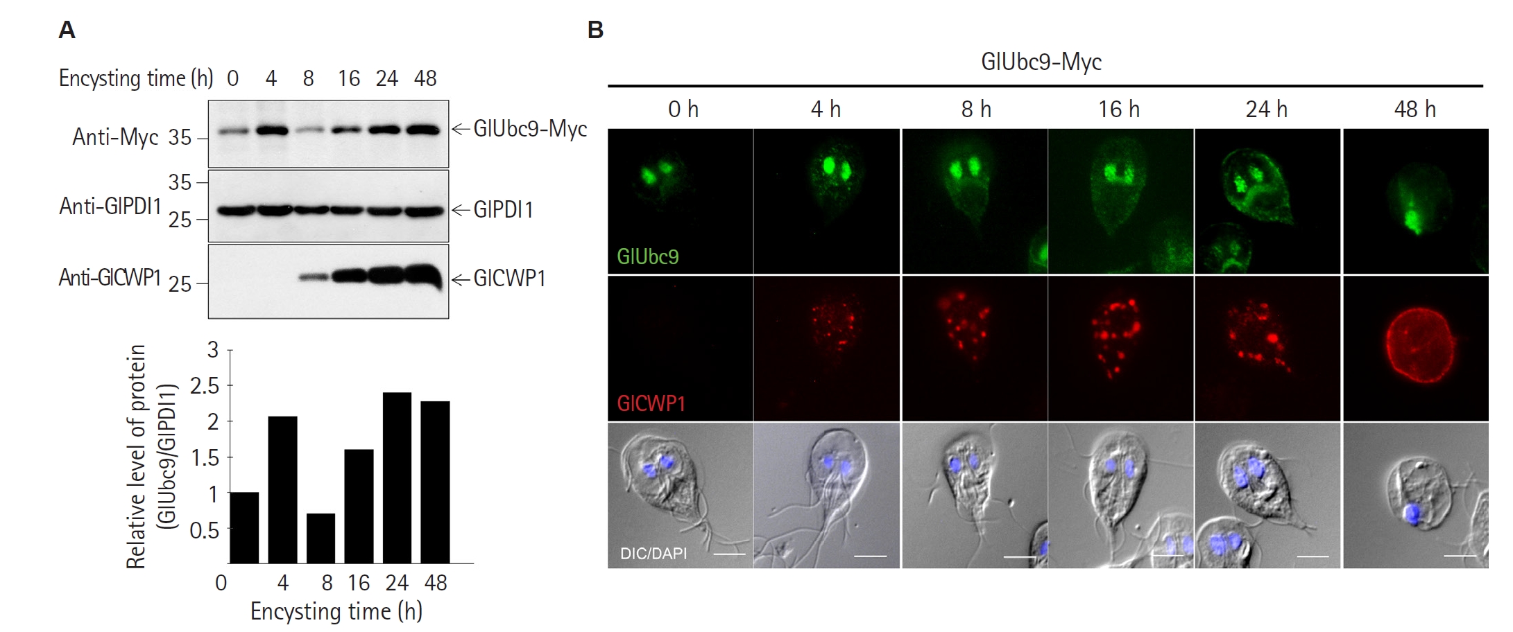
G. lamblia cells expressing Myc-tagged GlUbc9. GlUbc9-Myc was continuously expressed from 0 to 48 h post-induction, with protein levels generally increasing during encystation despite a transient decrease at 8 h post-induction (
Fig. 1A). Immunofluorescence analysis further showed that Myc-tagged GlUbc9 remained localized to the nucleus throughout encystation, from trophozoites (0 h) to encysting cells (4−24 h) and mature cysts (48 h) (
Fig. 1B). To monitor encystation progression, cells were co-stained with anti-GlCWP1 antibody to visualize ESVs. GlCWP1-positive ESVs first appeared as discrete puncta in the cytoplasm at 4 h post-induction, gradually increasing in number and size before becoming incorporated into the cyst wall by 48 h (
Fig. 1B) consistent with previous reports [
25]. These findings demonstrate that GlUbc9 is continuously expressed and maintains its nuclear localization throughout encystation, supporting a potential role for SUMOylation during cyst formation.
To determine whether SUMOylation is required for cyst formation, cyst formation efficiency was compared between GlUbc9 KD and control cells. As described previously [
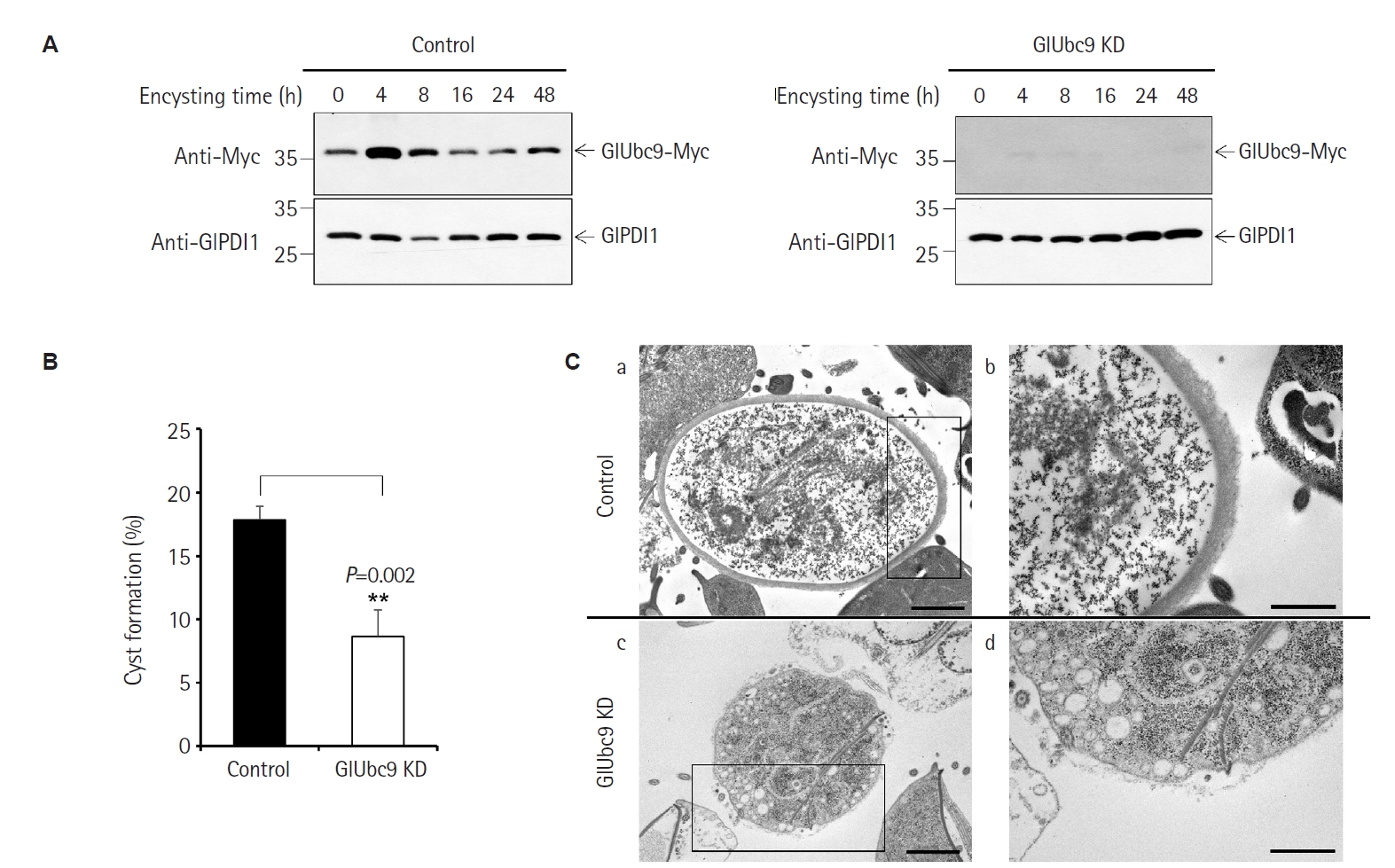
21], the GlUbc9 mRNA levels in CRISPRi-mediated GlUbc9 KD cells were reduced to approximately 15% of those in control cells. These cell lines were used throughout this study. Western blot analysis further confirmed that GlUbc9-Myc protein levels were markedly reduced in GlUbc9 KD cells compared with control cells throughout the encystation time course (
Fig. 2A).
The percentage of water-resistant cysts was significantly lower in GlUbc9 KD cells than in control cells (8.6% vs. 17.9%,
P=0.002) (
Fig. 2B). To evaluate cyst wall integrity, 48 h-encysting cells were examined by transmission electron microscopy. In control cells, mature cysts displayed a thick, well-defined, electron-dense cyst wall surrounding the cell (
Fig. 2Ca,
b). In contrast, cyst-like forms in GlUbc9 KD cells frequently exhibited a markedly thinner and loosely assembled cyst wall, while some lacked a discernible cyst wall altogether (
Fig. 2Cc,
d). These findings suggest that SUMOylation is required for efficient cyst formation and proper cyst wall assembly during
G. lamblia encystation.
The
Giardia cyst wall is composed of 3 cyst wall proteins (GlCWP1, GlCWP2, and GlCWP3) and a β-1,3-linked GalNAc carbohydrate homopolymer [
5,
26]. The abnormal cyst wall morphology observed in GlUbc9 KD cells by TEM (
Fig. 2Cc,
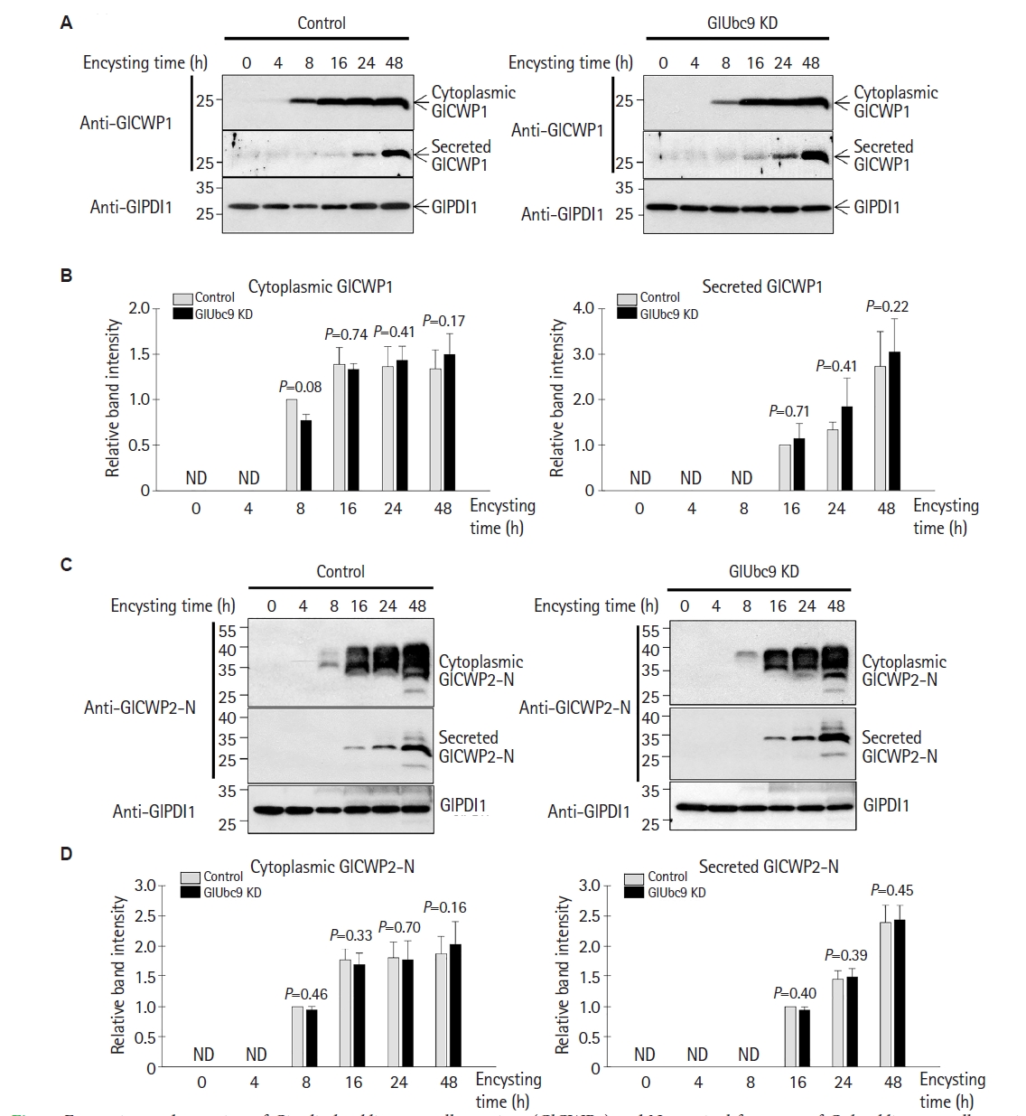
d) raised the possibility that cyst wall protein expression might be impaired. To investigate this possibility, the levels of GlCWP1 and GlCWP2 were examined in both cytoplasmic and secretory fractions during encystation. In control cells, cytoplasmic GlCWP1 was detectable from 8 h post-induction and remained present through 48 h, while secreted GlCWP1 appeared in the secretory fraction from 16 h onward (
Fig. 3A). Similar expression and secretion patterns were observed in control and GlUbc9 KD cells, and quantitative analysis showed no significant differences in cytoplasmic or secreted GlCWP1 levels between the 2 groups at any of the examined time points (
Fig. 3A,
B).
To assess GlCWP2 expression, an antibody specific to the N-terminal fragment of GlCWP2 (anti-GlCWP2-N) was generated (see Methods for details). To verify its specificity, recombinant GlCWP2 full-length (rGlCWP2-F), N-terminal (rGlCWP2-N), and C-terminal (rGlCWP2-C) proteins were expressed and analyzed by western blot. Anti-His antibody detected all 3 recombinant proteins, while anti-GlCWP2-N antibody reacted specifically with rGlCWP2-F and rGlCWP2-N but not rGlCWP2-C (
Supplementary Fig. S1). Using the anti-GlCWP2-N antibody, cytoplasmic GlCWP2 was detected from 8 h post-induction in both control and GlUbc9 KD cells, with secreted GlCWP2-N appearing from 16 h onward (
Fig. 3C). Quantitative analysis showed no significant differences in cytoplasmic or secreted GlCWP2-N levels between control and GlUbc9 KD cells at the examined time points (
Fig. 3D).
Since GlCWP2 undergoes proteolytic cleavage during encystation to generate an approximately 26 kDa GlCWP2-N and a 13 kDa GlCWP2-C [
11,
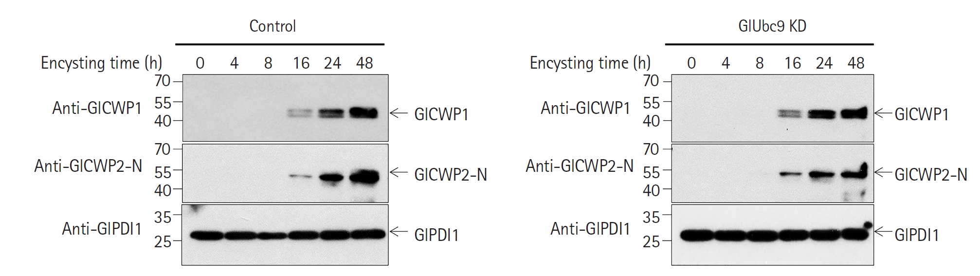
27], we next examined whether GlUbc9 KD affected this processing or the subsequent association of GlCWP2-N with GlCWP1 under non-reducing conditions. Immunoblotting with anti-GlCWP1 and anti-GlCWP2-N antibodies detected a band of approximately 50 kDa in both control and GlUbc9 KD cells throughout encystation (
Fig. 4). The molecular size of this band, together with its detection by both antibodies, is consistent with the formation of a GlCWP1–GlCWP2-N complex. No apparent difference in the abundance of this band was observed between control and GlUbc9 KD cells, suggesting that GlUbc9 KD does not impair GlCWP2 processing or its association with GlCWP1 during encystation. Equal protein loading of secretory fractions was confirmed by Coomassie staining, with overall protein profiles comparable between control and GlUbc9 KD cells (
Supplementary Fig. S2). Taken together, these findings indicate that GlCWP expression and proteolytic processing are largely preserved in GlUbc9 KD cells during encystation.
Despite the largely conserved expression and proteolytic processing of GlCWPs, cyst wall formation was impaired in GlUbc9 KD cells. We therefore hypothesized that intracellular trafficking and secretion of cyst wall components might be impaired. GlCWPs are transported to the cell surface via ESVs, which begin to form following GlCWP synthesis during encystation [
8,
27]. GalNAc homopolymer has been reported to be secreted via ECVs, which are labeled by DBA lectin [
9].
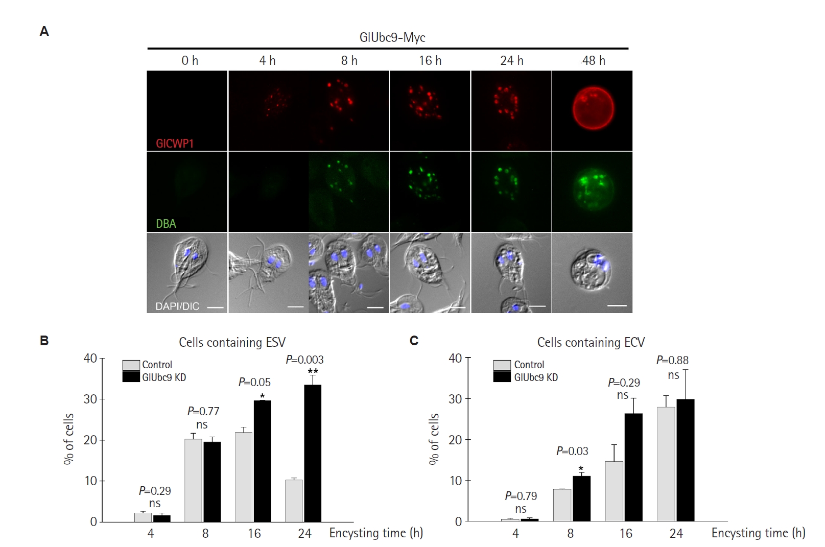
To investigate whether SUMOylation affects the formation of ESVs and ECVs, co-immunofluorescence staining with anti-GlCWP1 antibody (ESV marker) and FITC-conjugated DBA lectin (ECV marker) was performed. In
G. lamblia cells expressing GlUbc9-Myc, GlCWP1-positive ESVs were first detected at 4 h post-induction and increased through 24 h, while DBA-positive ECVs appeared from 8 h onward, consistent with the reported sequential formation of these 2 distinct vesicle populations during encystation (
Fig. 5A).
Quantitative analysis of ESV-containing cells across 4 independent experiments revealed that the percentage of cells containing ESVs was significantly higher in GlUbc9 KD cells than in control cells at 16 h (
P=0.05) and 24 h post-induction (
P=0.003), while no significant difference was observed at 4 h (
P=0.29) or 8 h (
P=0.77) (
Fig. 5B). These results suggest that ESVs accumulate intracellularly in GlUbc9 KD cells during the late stages of encystation.
In contrast, the percentage of ECV-containing cells was higher in GlUbc9 KD cells only at 8 h post-induction (
P=0.03), with no differences observed at 4 h (
P=0.79), 16 h (
P=0.29), or 24 h (
P=0.88) (
Fig. 5C), indicating that GlUbc9 KD did not substantially affect the overall formation of ECVs during encystation.
To further examine whether GalNAc carbohydrate secretion was affected by GlUbc9 KD, secretory fractions from control and GlUbc9 KD encysting cells were analyzed by western blot using biotinylated DBA lectin. Densitometric analysis of representative DBA-reactive bands at approximately 20 and 130 kDa showed similar secretion patterns between control and GlUbc9 KD cells throughout the encystation time course (
Supplementary Fig. S3). Equal protein loading of secretory fractions was confirmed by Coomassie staining (
Supplementary Fig. S2). No apparent alteration in GalNAc-containing glycoconjugates secretion was observed in GlUbc9 KD cells. Collectively, these findings indicate that depletion of GlUbc9 leads to intracellular accumulation of ESVs without markedly altering overall ECV formation or the secretion of GalNAc-containing glycoconjugates, suggesting that SUMOylation may contribute to ESV maturation and/or dynamics during
G. lamblia encystation.
Discussion
Encystation is a differentiation process that involves extensive cellular remodeling required for cyst wall biogenesis in
G. lamblia. Since this process requires coordinated regulation of multiple cellular events, post-translational modifications are likely to play important regulatory roles during parasite differentiation [
1]. SUMOylation is a reversible post-translational modification that regulates diverse cellular responses to environmental stress and a variety of biological processes, including cell cycle progression, protein trafficking, and cellular differentiation in eukaryotes [
15]. In
G. lamblia, we previously demonstrated that depletion of the SUMO-conjugating enzyme GlUbc9 impaired cell cycle progression and cell morphology in trophozoites [
21], suggesting that SUMOylation is required for normal parasite growth. However, whether SUMOylation contributes to the differentiation process during encystation has remained unknown. Therefore, we investigated the role of SUMOylation during encystation using a CRISPRi-mediated GlUbc9 (GL50803_24068) KD cell line.
GlUbc9 was continuously expressed throughout in vitro encystation, with protein levels gradually increasing over time (
Fig. 1A). This expression pattern suggests that SUMOylation may function not only during
Giardia trophozoite growth but also throughout encystation and may contribute to the regulation of this differentiation process. In addition, GlUbc9 maintained its nuclear localization throughout encystation (
Fig. 1B), indicating that the SUMO-conjugation machinery remains available throughout this process. These observations prompted us to investigate whether SUMOylation plays a functional role during
Giardia encystation.
GlUbc9 KD cells, in which GlUbc9-Myc expression was markedly reduced, produced significantly fewer water-resistant cysts than control cells (
Fig. 2B). TEM analysis further revealed that most cyst-like forms lacked a recognizable cyst wall or contained poorly organized cyst wall material (
Fig. 2C). We previously showed that GlUbc9 depletion impairs cell cycle progression and cell morphology in trophozoites [
21]; therefore, we cannot exclude the possibility that reduced cellular fitness may have contributed to the decreased cyst formation efficiency observed in GlUbc9 KD cells. However, previous studies have demonstrated that perturbation of the ubiquitin-proteasome system, another post-translational regulatory pathway, impairs GlCWP1 expression and efficient cyst formation during
Giardia encystation [
28]. These observations suggest that post-translational regulatory mechanisms play important roles during
Giardia encystation. In this context, the absence of a properly assembled cyst wall in GlUbc9 KD cells suggests that SUMOylation contributes to one or more steps involved in cyst wall biogenesis.
To determine which stage of cyst wall biogenesis was affected by GlUbc9 depletion, we examined the synthesis and maturation of the major cyst wall components. We first investigated whether the defective cyst wall phenotype resulted from impaired GlCWP expression. Quantitative analysis showed no significant differences in the cytoplasmic expression levels of GlCWP1 and GlCWP2-N between GlUbc9 KD and control cells at any of the examined time points (
Fig. 3), indicating that GlUbc9 depletion does not substantially affect GlCWP expression during encystation. We further found that proteolytic processing of GlCWP2 and the formation of the GlCWP1−GlCWP2-N complex under non-reducing conditions were largely preserved following GlUbc9 depletion (
Fig. 4). Although GlCWP3 could not be directly examined because a specific antibody was not successfully generated, the normal processing of GlCWP2 together with the preserved formation of the GlCWP1−GlCWP2-N complex suggests that the major events involved in cyst wall protein maturation remain intact. Furthermore, densitometric analysis of representative DBA-reactive bands showed similar secretion patterns of GalNAc-containing glycoconjugates between GlUbc9 KD and control cells (
Supplementary Fig. S3). Since GalNAc synthesis and secretion are essential for proper cyst wall formation [
10,
29], these findings suggest that GalNAc secretory pathway is largely preserved following GlUbc9 depletion. Collectively, these observations indicate that the cyst wall defects in GlUbc9 KD cells are unlikely to result from impaired synthesis or post-translational maturation of cyst wall components.
Since the major biosynthetic processes required for cyst wall formation appeared to be unchanged, we next examined ESV formation and dynamics during encystation. ESVs in
Giardia are Golgi-like organelles that form de novo during encystation in close association with GlCWP synthesis [
8]. Consistent with this role, GlUbc9 depletion did not affect the initial formation of ESVs at 4 h post-induction but resulted in marked intracellular accumulation of ESVs during the late stages of encystation (16–24 h post-induction) (
Fig. 5B). These findings suggest that SUMOylation is dispensable for the early formation of ESVs but may contribute to their subsequent maturation and/or dynamics.
Previous studies have shown that several small GTPases, including Sar1, Arf1, and Rab1, regulate ESV biogenesis and trafficking during encystation [
7,
8]. Among these, expression of a dominant-negative Arf1 mutant impairs secretion of cyst wall material and results in the formation of non-water-resistant naked cysts despite normal ESV formation [
8]. The phenotype observed in GlUbc9 KD cells therefore shares features with perturbation of late-stage ESV maturation rather than defective initial ESV formation.
However, despite intracellular accumulation of ESVs, quantitative analysis showed no significant differences in secreted GlCWP1 or GlCWP2-N levels between GlUbc9 KD and control cells (
Fig. 3). Thus, GlUbc9 depletion does not appear to completely block GlCWP secretion. One possible explanation is that GlUbc9 depletion alters the timing or dynamics of ESV maturation, resulting in delayed rather than abolished secretion of cyst wall material, which may be insufficient to support proper cyst wall assembly.
Only a limited number of SUMO substrates have been identified in
G. lamblia, including α-tubulin [
30], arginine deiminase [
31,
32], 2 centrin proteins [
21], never in mitosis A-related kinase, aminoacyl-histidine dipeptidase, and protein disulfide isomerase 2 [
22]. Because the phenotype of GlUbc9 KD cells resembles that of cells expressing inactive Arf1 (GL50803_7789), we considered Arf1 to be a potential SUMO substrate during encystation. To explore this possibility, we analyzed the
Giardia Arf1 amino acid sequence using the GPS-SUMO 1.0 prediction tool [
33], which identified 2 putative SUMOylation sites at lysine residues K144 and K190. Although these predictions do not demonstrate that Arf1 is directly SUMOylated, they support the possibility that Arf1 may represent a candidate SUMO substrate potentially involved in ESV maturation or dynamics. Experimental validation, such as co-immunoprecipitation using epitope-tagged Arf1, will be required to determine whether Arf1 is directly SUMOylated during encystation.
Taken together, our findings indicate that SUMOylation is required for G. lamblia encystation and suggest that its role is associated with the timely maturation and/or dynamics of ESVs rather than with GlCWP expression or proteolytic processing. These findings reveal a previously unrecognized role for SUMOylation during Giardia encystation and provide a foundation for future studies aimed at elucidating the molecular mechanisms by which SUMOylation coordinates infectious cyst formation in Giardia.
Notes
-
Data availability
Raw data are available upon request from the corresponding author.
-
Author contributions
Conceptualization: Kim J, Park SJ. Data curation: Shin MY, Kim J. Formal analysis: Kim J. Funding acquisition: Kim J, Park SJ. Investigation: Yeo HR, Shin MY, Kim J. Methodology: Kim J, Park SJ. Project administration: Park SJ. Supervision: Kim J. Validation: Shin MY. Visualization: Yeo HR, Kim J. Writing – original draft: Yeo HR, Kim J. Writing – review & editing: Kim J, Park SJ.
-
Conflict of interest
Juri Kim and Soon-Jung Park serve as editors of Parasites, Hosts and Diseases but had no involvement in the decision to publish this article. No other potential conflicts of interest relevant to this study were reported.
-
Funding
This work was supported by the National Research Foundation of Korea (NRF) grant funded by the Korea government (MSIT) (grant No. RS-2023-NR076338 to SJ Park and grant No. RS-2025-24683462 to J Kim).
Supplementary information
Fig. 1.
Giardia lamblia SUMO-conjugating enzyme Ubc9 (GlUbc9) is continuously expressed during G. lamblia encystation. (A) Western blot analysis of GlUbc9-Myc expression during encystation. Lysates from G. lamblia cells expressing Myc-tagged GlUbc9 were prepared at the indicated time points (0–48 h post-induction) and analyzed using an anti-Myc antibody. G. lamblia protein disulfide isomerase 1 (GlPDI1) was used as a loading control. The bar graph shows the relative GlUbc9-Myc protein levels normalized to GlPDI1. G. lamblia cyst wall protein 1 (GlCWP1) expression is shown as a marker of encystation. (B) Representative immunofluorescence images showing the localization of GlUbc9-Myc (green, anti-Myc) and GlCWP1 (red, anti-GlCWP1) in G. lamblia cells at the indicated time points during encystation. Nuclei were stained with 4′,6-diamidino-2-phenylindole (DAPI, blue). DIC, differential interference contrast. Scale bar=5 μm.
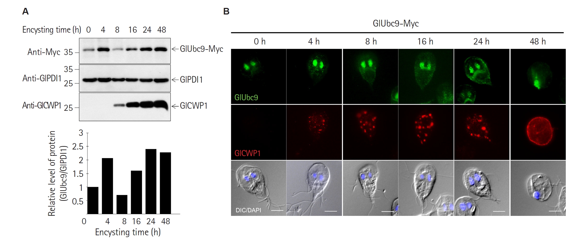
Fig. 2.
Giardia lamblia SUMO-conjugating enzyme Ubc9 (GlUbc9) knockdown (KD) reduces cyst formation efficiency in G. lamblia. (A) Western blot analysis confirming CRISPRi-mediated knockdown of GlUbc9-Myc in control and GlUbc9 KD cells at the indicated time points (0–48 h post-induction). Membranes were probed with an anti-Myc antibody. G. lamblia protein disulfide isomerase 1 (GlPDI1) was used as a loading control. (B) Cyst formation efficiency of control and GlUbc9 KD cells at 48 h post-induction. Water-resistant cysts were quantified using a hemocytometer. Data are presented as the mean±SE from 3 independent experiments. **P<0.01. (C) Representative transmission electron microscopy images of control (a, b) and GlUbc9 KD (c, d) cells after 48 h of encystation. Panels b and d show higher-magnification views of the boxed regions in panels a and c, respectively. Scale bar=2 μm.
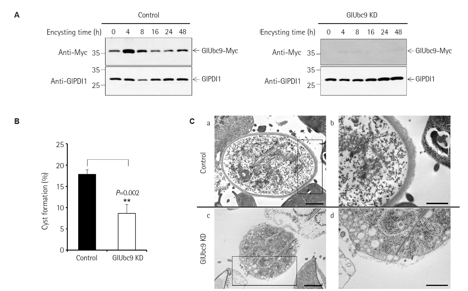
Fig. 3.Expression and secretion of Giardia lamblia cyst wall protein 1 (GlCWP1) and N-terminal fragment of G. lamblia cyst wall protein 2 (GlCWP2-N) in control and G. lamblia SUMO-conjugating enzyme Ubc9 (GlUbc9) knockdown (KD) cells during encystation. (A) Western blot analysis of cytoplasmic and secretory fractions from control and GlUbc9 KD encysting cells probed with an anti-GlCWP1 antibody at the indicated time points (0–48 h post-induction). GlPDI1 was used as a loading control for cytoplasmic fractions. (B) Quantification of cytoplasmic GlCWP1 (left) and secreted GlCWP1 (right) band intensities. (C) Western blot analysis of cytoplasmic and secretory fractions from control and GlUbc9 KD encysting cells probed with anti-GlCWP2-N antibody at the indicated time points (0–48 h post-induction). GlPDI1 was used as a loading control for cytoplasmic fractions. See Supplementary Fig. S1 for characterization of the anti-GlCWP2-N antibody. (D) Quantification of cytoplasmic GlCWP2-N (left) and secreted GlCWP2-N (right) band intensities. For densitometric analysis, band intensities were normalized to GlPDI1 for cytoplasmic fractions or to a Coomassie-stained loading control band for secretory fractions. For graphical presentation, the normalized value of control cells at the first detectable time point (8 h for cytoplasmic fractions; 16 h for secretory fractions) in each independent experiment was set to 1.0. For all graphs, data are presented as mean±SE from 3 independent experiments. Statistical significance was determined by paired Student t-test. ND, not detected.
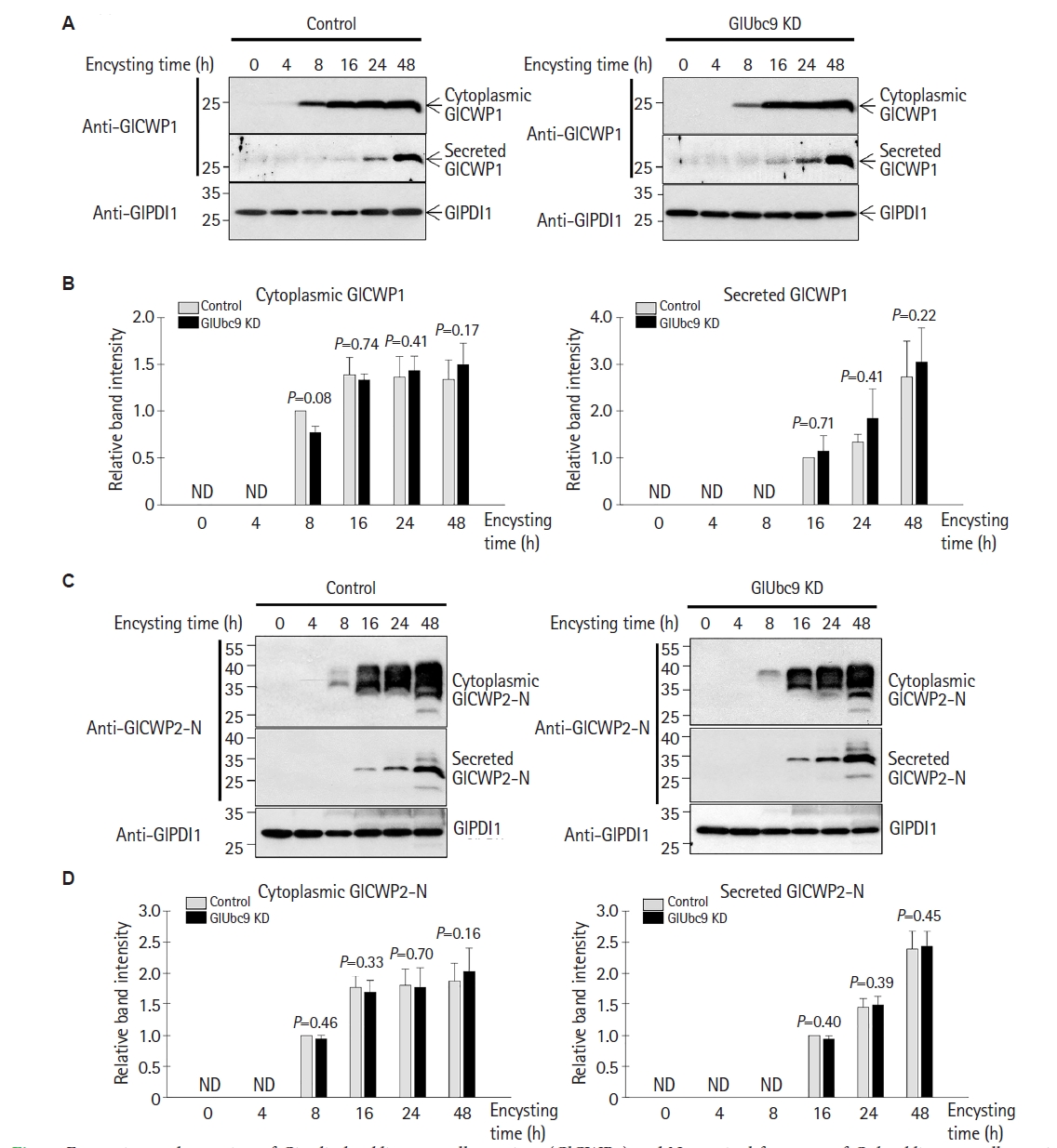
Fig. 4.Detection of a Giardia lamblia cyst wall protein 1 (GlCWP1)−N-terminal fragment of G. lamblia cyst wall protein 2 (GlCWP2-N) complex under non-reducing conditions. Western blot analysis of cytoplasmic fractions from control and G. lamblia SUMO-conjugating enzyme Ubc9 (GlUbc9) knockdown (KD) encysting cells collected at the indicated time points (0–48 h post-induction). Samples were analyzed under non-reducing conditions using anti-GlCWP1 and anti-GlCWP2-N antibodies. GlPDI1 was used as a loading control. A representative blot from 3 independent experiments is shown.
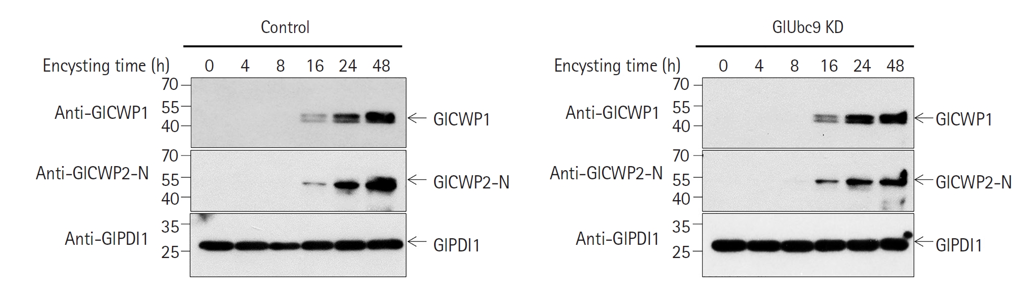
Fig. 5.Encystation-specific vesicle (ESV) and encystation-specific carbohydrate-positive vesicle (ECV) formation in Giardia lamblia SUMO-conjugating enzyme Ubc9 (GlUbc9) knockdown (KD) during encystation. (A) Representative immunofluorescence images of GlUbc9-Myc-expressing G. lamblia cells at the indicated time points (0–48 h post-induction) stained with an anti–Giardia lamblia cyst wall protein 1 (GlCWP1) antibody (red, ESV marker) and fluorescein isothiocyanate-conjugated Dolichos biflorus agglutinin (DBA) lectin (green, ECV marker). Nuclei were stained with 4′,6-diamidino-2-phenylindole (DAPI, blue). DIC, differential interference contrast. Scale bar=5 μm. (B) Quantification of cells containing ESVs (GlCWP1-positive) in control and GlUbc9 KD cells at 4, 8, 16, and 24 h post-induction. (C) Quantification of cells containing ECVs (DBA-positive) in control and GlUbc9 KD cells at 4, 8, 16, and 24 h post-induction. For panels (B) and (C), more than 200 cells were counted per sample in each of 4 independent experiments. Data are presented as the mean±SE. *P<0.05, **P<0.01. ns, not significant.
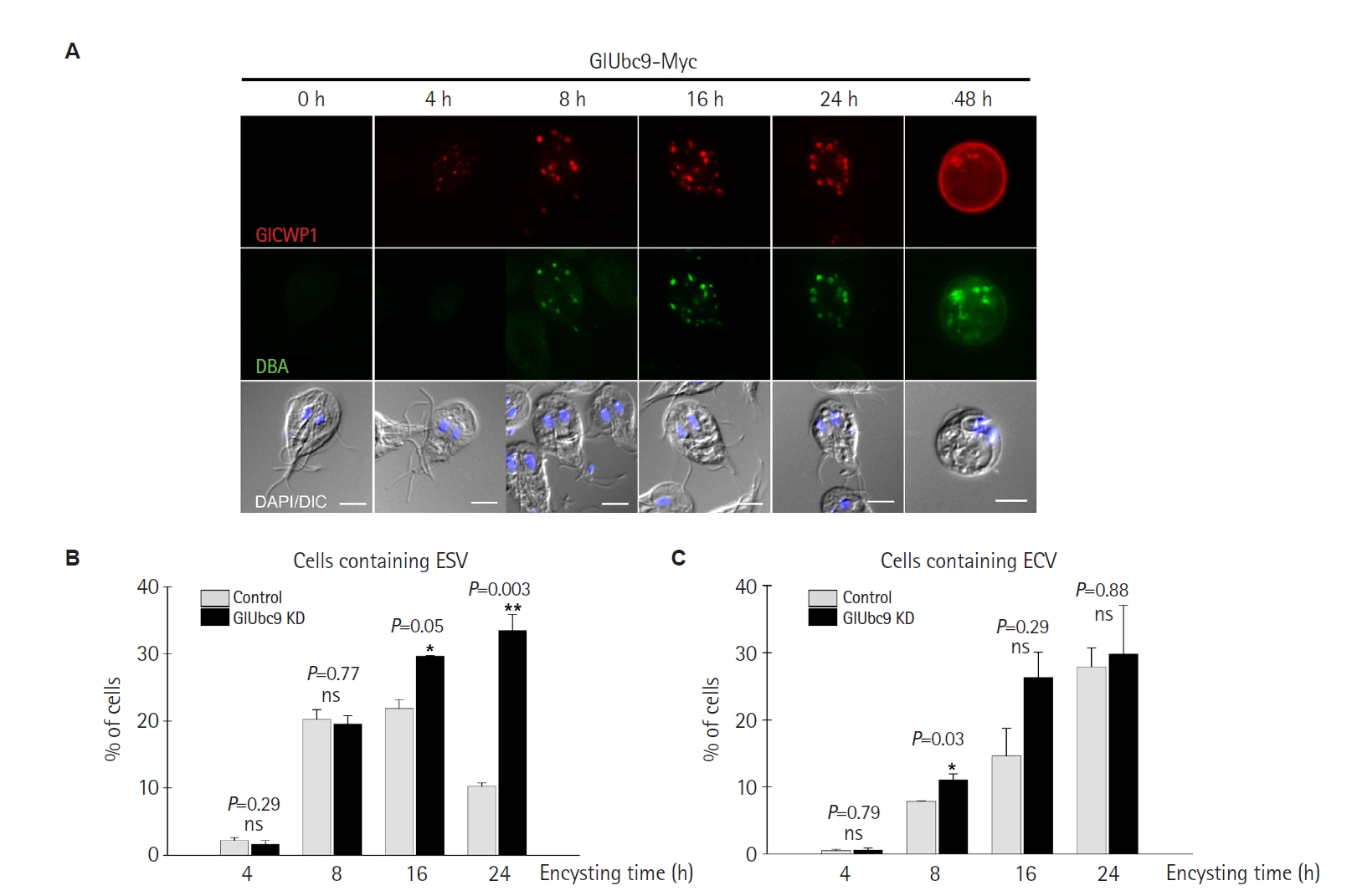
Table 1.Primers used in this study
Table 1.
|
Name |
Nucleotide sequence (5'-3')a
|
|
rCWP2-F-F |
CATGCCATGGGCATGATCGCAGCCCTTGT |
|
rCWP2-F-R |
GTTAGCGGCCGCCCTTCTGCGGACAATAG |
|
rCWP2-N-F |
GCCGAATTCGATGATCGCAGCCCTTGTTCTAGG |
|
rCWP2-N-R |
ATAAGAATGCGGCCGCCGTCTTGCTAGATGACCTAACCA |
|
rCWP2-C-F |
GCCGAATTCGGCCTCAACGTCCCGCTCCA |
|
rCWP2-C-R |
ATAAGAATGCGGCCGCTCTTCTGCGGACAATAGGCTTG |
Table 2.Strains and plasmids used in this study
Table 2.
|
Organism/plasmid |
Description |
Source/reference |
|
Giardia lamblia
|
|
|
|
ATCC 30957 |
Clinical isolate |
ATCC |
|
GlUbc9-Myc |
G. lamblia expressing C-terminal Myc-tagged GlUbc9 |
[21] |
|
GlUbc9 knockdown |
CRISPRi-mediated knockdown of GlUbc9 |
[21] |
|
Escherichia coli
|
|
|
|
DH5α |
supE44, ΔlacU169 (Φ80 lacZ ΔM15), hsdR17, recA1, endA1, gyrA96, thi-1, relA1
|
Invitrogen |
|
BL21 (DE3) |
F’, ompT, hsdSB(rB-mB-) gal, dcm (DE3)
|
Invitrogen |
|
Plasmids |
|
|
|
pET28b |
Expression vector for histidine-tagged protein, KanR
|
Novagen |
|
pET28-CWP2 |
pET28b, 1,089-bp encoding Giardia cwp2 gene (GiardiaDB: GL50803_5435) |
This study |
|
pET21b |
Expression vector for histidine-tagged protein, AmpR
|
Novagen |
|
pET21-CWP2-N |
pET21b, 912-bp encoding N-terminus of Giardia cwp2 gene (GiardiaDB: GL50803_5435) |
This study |
|
pET21-CWP2-C |
pET21b, 204-bp encoding C-terminus of Giardia cwp2 gene (GiardiaDB: GL50803_5435) |
This study |
References
- 1. Adam RD. Giardia duodenalis: biology and pathogenesis. Clin Microbiol Rev 2021;34:e0002419. https://doi.org/10.1128/CMR.00024-19
- 2. Bourli P, Eslahi AV, Tzoraki O, Karanis P. Waterborne transmission of protozoan parasites: a review of worldwide outbreaks: an update 2017-2022. J Water Health 2023;21:1421-47. https://doi.org/10.2166/wh.2023.094
- 3. Luján HD, Mowatt MR, Conrad JT, Bowers B, Nash TE. Identification of a novel Giardia lamblia cyst wall protein with leucine-rich repeats. Implications for secretory granule formation and protein assembly into the cyst wall. J Biol Chem 1995;270:29307-13. https://doi.org/10.1074/jbc.270.49.29307
- 4. Sun CH, McCaffery JM, Reiner DS, Gillin FD. Mining the Giardia lamblia genome for new cyst wall proteins. J Biol Chem 2003;278:21701-8. https://doi.org/10.1074/jbc.M302023200
- 5. Chatterjee A, Carpentieri A, Ratner DM, et al. Giardia cyst wall protein 1 is a lectin that binds to curled fibrils of the GalNAc homopolymer. PLoS Pathog 2010;6:e1001059. https://doi.org/10.1371/journal.ppat.1001059
- 6. Marti M, Hehl AB. Encystation-specific vesicles in Giardia: a primordial Golgi or just another secretory compartment? Trends Parasitol 2003;19:440-6. https://doi.org/10.1016/s1471-4922(03)00201-0
- 7. Faso C, Konrad C, Schraner EM, Hehl AB. Export of cyst wall material and Golgi organelle neogenesis in Giardia lamblia depend on endoplasmic reticulum exit sites. Cell Microbiol 2013;15:537-53. https://doi.org/10.1111/cmi.12054
- 8. Stefanic S, Morf L, Kulangara C, et al. Neogenesis and maturation of transient Golgi-like cisternae in a simple eukaryote. J Cell Sci 2009;122:2846-56. https://doi.org/10.1242/jcs.049411
- 9. Midlej V, Meinig I, de Souza W, Benchimol M. A new set of carbohydrate-positive vesicles in encysting Giardia lamblia. Protist 2013;164:261-71. https://doi.org/10.1016/j.protis.2012.11.001
- 10. Krtková J, Thomas EB, Alas GC, et al. Rac regulates Giardia lamblia encystation by coordinating cyst wall protein trafficking and secretion. mBio 2016;7:e01003-16. https://doi.org/10.1128/mBio.01003-16
- 11. Touz MC, Nores MJ, Slavin I, et al. The activity of a developmentally regulated cysteine proteinase is required for cyst wall formation in the primitive eukaryote Giardia lamblia. J Biol Chem 2002;277:8474-81. https://doi.org/10.1074/jbc.M110250200
- 12. DuBois KN, Abodeely M, Sakanari J, et al. Identification of the major cysteine protease of Giardia and its role in encystation. J Biol Chem 2008;283:18024-31. https://doi.org/10.1074/jbc.M802133200
- 13. Konrad C, Spycher C, Hehl AB. Selective condensation drives partitioning and sequential secretion of cyst wall proteins in differentiating Giardia lamblia. PLoS Pathog 2010;6:e1000835. https://doi.org/10.1371/journal.ppat.1000835
- 14. Geiss‐Friedlander R, Melchior F. Concepts in sumoylation: a decade on. Nat Rev Mol Cell Biol 2007;8:947-56. https://doi.org/10.1038/nrm2293
- 15. Vertegaal ACO. Signalling mechanisms and cellular functions of SUMO. Nat Rev Mol Cell Biol 2022;23:715-31. https://doi.org/10.1038/s41580-022-00500-y
- 16. Seufert W, Futcher B, Jentsch S. Role of a ubiquitin-conjugating enzyme in degradation of S- and M-phase cyclins. Nature 1995;373:78-81. https://doi.org/10.1038/373078a0
- 17. Jones D, Crowe E, Stevens TA, Candido EPM. Functional and phylogenetic analysis of the ubiquitylation system in Caenorhabditis elegans: ubiquitin-conjugating enzymes, ubiquitin-activating enzymes, and ubiquitin-like proteins. Genome Biol 2002;3:RESEARCH0002. https://doi.org/10.1186/gb-2001-3-1-research0002
- 18. Nacerddine K, Lehembre F, Bhaumik M, et al. The SUMO pathway is essential for nuclear integrity and chromosome segregation in mice. Dev Cell 2005;9:769-79. https://doi.org/10.1016/j.devcel.2005.10.007
- 19. Vranych CV, Merino MC, Zamponi N, Touz MC, Rópolo AS. SUMOylation in Giardia lamblia: a conserved post‐translational modification in one of the earliest divergent eukaryotes. Biomolecules 2012;2:312-30. https://doi.org/10.3390/biom2030312
- 20. Castellanos IC, Calvo EP, Wasserman M. A new gene inventory of the ubiquitin and ubiquitin‐like conjugation pathways in Giardia intestinalis. Mem Inst Oswaldo Cruz 2020;115:e190242. https://doi.org/10.1590/0074-02760190242
- 21. Yeo HR, Shin MY, Kim J, Park SJ. Two centrins and their posttranslational modification modulate the cell cycle of Giardia lamblia. Microbiologyopen 2025;14:e70038. https://doi.org/10.1002/mbo3.70038
- 22. Yeo HR, Shin MY, Kim J, Park SJ. Identification and confirmation of SUMOylation-modified proteins in Giardia trophozoites. Parasites Hosts Dis 2025;63:264-71. https://doi.org/10.3347/PHD.25023
- 23. Einarsson E, Troell K, Hoeppner MP, et al. Coordinated changes in gene expression throughout encystation of Giardia intestinalis. PLoS Negl Trop Dis 2016;10:e0004571. https://doi.org/10.1371/journal.pntd.0004571
- 24. Kim J, Park SJ. Role of gamma‐giardin in ventral disc formation of Giardia lamblia. Parasit Vectors 2019;12:227. https://doi.org/10.1186/s13071-019-3478-8
- 25. Kim J, Park EA, Shin MY, Park SJ. Identification of target genes regulated by encystation-induced transcription factor Myb2 using knockout mutagenesis in Giardia lamblia. Parasit Vectors 2022;15:360. https://doi.org/10.1186/s13071-022-05489-z
- 26. Jarroll EL, Macechko PT, Steimle PA, et al. Regulation of carbohydrate metabolism during Giardia encystment. J Eukaryot Microbiol 2001;48:22-6. https://doi.org/10.1111/j.1550-7408.2001.tb00412.x
- 27. Gottig N, Elías EV, Quiroga R, et al. Active and passive mechanisms drive secretory granule biogenesis during differentiation of the intestinal parasite Giardia lamblia. J Biol Chem 2006;281:18156-66. https://doi.org/10.1074/jbc.M602081200
- 28. Alvarado ME, Chaparro-Gutiérrez JJ, Calvo EP, et al. Activity of the Giardia intestinalis proteasome during encystation and its connection with the expression of the cyst wall protein 1 (CWP1). Acta Trop 2022;225:106183. https://doi.org/10.1016/j.actatropica.2021.106183
- 29. Sener K, Shen Z, Newburg DS, Jarroll EL. Amino sugar phosphate levels in Giardia change during cyst wall formation. Microbiology (Reading) 2004;150:1225-30. https://doi.org/10.1099/mic.0.26898-0
- 30. Di Genova BM, da Silva RC, da Cunha JPC, et al. Protein SUMOylation is involved in cell‐cycle progression and cell morphology in Giardia lamblia. J Eukaryot Microbiol 2017;64:491-503. https://doi.org/10.1111/jeu.12386
- 31. Touz MC, Rópolo AS, Rivero MR, et al. Arginine deiminase has multiple regulatory roles in the biology of Giardia lamblia. J Cell Sci 2008;121:2930-8. https://doi.org/10.1242/jcs.026963
- 32. Vranych CV, Rivero MR, Merino MC, et al. SUMOylation and deimination of proteins: two epigenetic modifications involved in Giardia encystation. Biochim Biophys Acta 2014;1843:1805-17. https://doi.org/10.1016/j.bbamcr.2014.04.014
- 33. Zhao Q, Xie Y, Zheng Y, et al. GPS-SUMO: a tool for the prediction of sumoylation sites and SUMO-interaction motifs. Nucleic Acids Res 2014;42:W325-30. https://doi.org/10.1093/nar/gku383